01/12/2017 09:00 AM EST

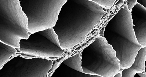
Over the past few years, my blog has highlighted winners from the annual BioArt contest sponsored by the Federation of American Societies for Experimental Biology (FASEB). So, let’s keep a good thing going with one of the amazing scientific images that captured top honors in FASEB’s latest competition: a scanning electron micrograph of the hamstring […]
Snapshots of Life: An Elegant Design
Credit: David Sleboda and Thomas Roberts, Brown University, Providence, RI
Over the past few years, my blog has highlighted winners from the annual BioArt contest sponsored by the Federation of American Societies for Experimental Biology (FASEB). So, let’s keep a good thing going with one of the amazing scientific images that captured top honors in FASEB’s latest competition: a scanning electron micrograph of the hamstring muscle of a bullfrog.
That’s right, a bullfrog, For decades, researchers have used the American bullfrog, Rana catesbeiana, as a model for studying the physiology and biomechanics of skeletal muscles. My own early work with electron microscopy, as a student at Yale in the 1970s, was devoted to producing images from this very tissue. Thanks to its disproportionately large skeletal muscles, this common amphibian has played a critical role in helping to build the knowledge base for understanding how these muscles work in other organisms, including humans.
Revealed in this picture is the intricate matrix of connective tissue that holds together the frog’s hamstring muscle, with the muscle fibers themselves having been digested away with chemicals. And running diagonally, from lower left to upper right, you can see a band of fibrils made up of a key structural protein called collagen.
The image is the work of David Sleboda, a graduate student in the lab of Thomas Roberts at Brown University, Providence, RI. Sleboda studies muscle physiology, with a focus on how muscles specialize to perform different tasks. A classic example is one that joggers probably know well. Certain leg muscles act as motors to propel the body uphill, while others act as brakes to slow the downward descent.
Sleboda thinks muscle specialization may be linked to structural variations in the connective tissue that surrounds muscle fibers. It’s an intriguing idea, and one that’s been surprisingly understudied. When Sleboda decided to survey the connective tissue in various muscles, his first stop was the bullfrog model.
But Sleboda soon realized that no one had ever checked to confirm that bullfrogs actually have connective tissue around their skeletal muscle fibers. That task suddenly fell to him, and it presented a major technical challenge. Sleboda had no background in imaging. Neither did anyone in the lab. Undeterred, he spent several months testing out various imaging techniques before settling on scanning electron microscopy (SEM) for its unsurpassed resolution.
SEM involves bombarding tissue with beams of electrons rather than light, and it can magnify the world inside of our cells up to 10 million times. But it isn’t easy to master. Sleboda buckled down last summer, diving into the methods sections of a few select SEM research papers and turning to the knowledgeable minds at Brown’s bioimaging center for advice. By the end of the summer—and after lots of trial and error—Sleboda was able to snap a number of gems, including this one.
And he’s not stopping there. Sleboda continues to use SEM to generate informative images of the connective tissue in many different types of muscles and animals. Although his results have yet to be published, this sneak peek suggests lots of fascinating data still to come.
Links:
Healthy Muscles Matter (National Institute of Arthritis and Musculoskeletal and Skin Diseases/NIH)
Amphibians as animal models for laboratory research in physiology. Burggren WW, Warburton S. ILAR J. 2007;48(3):260-269.
Roberts Lab (Brown University, Providence, RI)
BioArt (Federation of American Societies for Experimental Biology, Bethesda, MD)
NIH Support: National Institute of Arthritis and Musculoskeletal and Skin Diseases


.png)


No hay comentarios:
Publicar un comentario