Chasing Fireflies—and Better Cellular Imaging Techniques

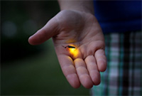
Firefly. Credit: Stock photo.
The yellow-green glow from this summer’s fireflies teased my kids across the yard. Max and Stella zigzagged the grass, occasionally jumping into the air to cup a firefly in their hands and then proudly shouting, “I got one!”
Chasing fireflies on a summer night is a childhood rite of passage for many, including Nathan Shaner who grew up in New Jersey. “It was one of my favorite things about summer,” he recalls. “I’d catch them with my hands—I’d never jar them.”
Today, Shaner studies the science of bioluminescence, which gives fireflies and many other organisms the natural ability to emit light. His goal is to make bright bioluminescent tags that he and other scientists can use to study living cells in greater detail. “There’s this very beautiful thing that evolved in nature, and we can use it to enable new discoveries,” he says.
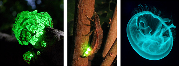
Thousands of organisms glow as a way to communicate, spook predators, lure prey or attract mates. There are a few terrestial examples, such as fireflies, glowworm insect larvae and foxfire fungi, and many more acquatic ones, including types of marine plankton, fish, jellyfish, shrimp, squid and sea urchins. One research team estimated nearly three quarters of sea life have bioluminescent capabilities.

Bioluminescence is common across the tree of life (left to right): Panellus Stipticus (foxfire fungi); Lampryis noctiluca (glowworm insect); Aurelia Aurita (moon jellyfish). Credit: Wikimedia Commons, Ylem; Wikimedia Commons, Wofl; stock photo.
Every studied case of bioluminescence involves oxygen, a light-emitting pigment called luciferin and a protein called luciferase. Luciferase encourages the pigment’s reaction with oxygen, releasing energy in the form of light. Although many bioluminescent creatures have their own form of luciferase, they share just a handful of luciferins. For example, the luciferin called coelenterazine is found in many aquatic organisms.
Shaner mainly works with coelenterazine in his California-based lab at the Scintillon Institute  . It’s his “favorite” luciferin, he says, for a variety of reasons. The brightest luciferases that use coelenterazine are half the size of green fluorescent protein, another natural light-emitting molecule scientists have harnessed for biological imaging. The small size, says Shaner, means these luciferases could be good for tagging proteins in a biological sample. Firefly luciferase, by comparison, is bigger and has slower reaction times.
Shaner has spent nearly a decade figuring out how aquatic creatures make coelenterazine and dissecting its interactions with luciferases. Knowing these details has helped him and his team create bioluminescent reactions that glow brighter and longer. Coelenterazine has just two amino acid building blocks, raising hopes that scientists may be able to coax other organisms into synthesizing it on their own.

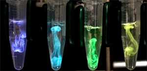
Details about the basic biology and chemistry of the ingredients that produce bioluminescence are allowing scientists to harness it as an imaging tool. Credit: Nathan Shaner, Scintillon Institute.
This picture, which Shaner took with his phone, shows four tubes containing the ingredients that produce bioluminescence. In each tube, coelenterazine appears buoyant, swirling as it interacts with luciferase. Luciferase alone emits a purple color. It emits other colors, as shown in the three tubes on the right, when it’s attached to different fluorescent proteins. The reactions captured here lasted up to 10 minutes, but Shaner says a continuous shot of luciferin could produce an inextinguishable glow.
The ability to use bioluminescence to image live cells and their parts offers several advantages over existing techniques. Bioluminescence imaging, unlike fluorescence imaging, does not require the application of external light. “The intensity of light in fluorescence imaging is enormous and is going to have an impact on the sample,” says Shaner, explaining that the light can kill or damage molecules in the cell. “We want to observe cells in their most pristine state.” The external light also can produce fluorescence arising from the natural contents of the cell, diminshing the contrast of the fluorescent signal from the parts of interest.
Although bioluminescence imaging provides less phototoxicity and better contrast, it is not nearly as bright as fluorescence imaging. The brightest luciferases emit about four photons per second. To be most useful as imaging tools, Shaner estimates they’ll need to emit up to 1,000 photons per second. Another drawback Shaner is working to overcome: Turning bioluminescence on and off, which would allow cell parts to light up as others go dark.
“Fluorescent proteins changed science so much when they were discovered,” says Shaner, who trained in the lab of fluorescent protein pioneer and Nobel laureate Roger Tsien. “I’m excited about the kinds of biological questions scientists may be able to answer with bioluminescence imaging.”
In the meantime, Max and Stella will continue to chase fireflies and, like Shaner, admire beauty in nature.


No hay comentarios:
Publicar un comentario