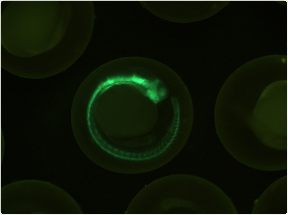
Fluorescent zebrafish genes provide clues about neuroblastoma
Researchers have created transgenic zebrafish that could help them understand how the third most common pediatric cancer in the U.S develops.
 Image Credit: lostkabab / Shutterstock
Neurobiologist and cancer researcher Rosa Uribe from Rice University in Houston, Texas, created the fish with colleagues from the University of Illinois and the California Institute of Technology.
As reported in the journal Genesis, the zebrafish produce tags that fluoresce in different colors depending on how a gene called SOX10 is behaving.
The gene is active in stem cells called neural crest cells. Observing how those cells migrate and transition could improve our understanding about neuroblastoma.
Using a time-lapse movie to track the cells, Uribe says: “You can see that a lot of them are transitioning. Some of them are dividing there, there, there. And then they turn off the green, which means they're done dividing."
The scientists would now like to understand why the cells stop dividing. SOX10 is one of twenty types of SOX proteins, all of which control rapid cell division in growing embryos.
These proteins are also often active in cancer cells and finding out what it is that switches SOX10 “off” in neural crest cells could help researchers develop treatments for cancers where SOX proteins are involved.
EMT is key to embryonic development as it enables cells to become more like stem cells and to form new tissues. However, studies have also shown that EMT is the switch used by cancer cells to spread or “metastasise.”
Being able to track these neural crest cells from the first moment they form through to once they have finished migrating is one important way to learn more about them.
Uribe and colleagues also intend to mix and match genes in new types of zebrafish to see what happens when cells produce too much or too little of a particular protein.


No hay comentarios:
Publicar un comentario