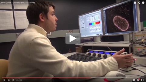
LabTV: Curious about Microscopy
Growing up amid the potato and corn fields of western New York state, Jordan Myers got a firsthand look at what it was like to work as a farmer, a homebuilder, even a chimney sweep. But it was television—specifically, “Bill Nye the Science Guy” and “The Magic School Bus”—that introduced him to what would become his future career: science.
Propelled by his curiosity about how living things work, Myers left his hometown of Savannah to attend New York’s Rochester Institute of Technology, where he earned an undergraduate degree in biotechnology, and then headed off to pursue advanced degrees in cell biology at Yale School of Medicine, New Haven, CT. There, as you’ll see in this LabTV profile, he’s trying to develop light microscopy techniques [1,2] to view the cell’s nuclear envelope at nanometer (nm) resolution—a major challenge when one considers that a red blood cell measures about 7,000 nm in diameter.
Working with colleagues in the labs of Charles Patrick Lusk and Joerg Bewersdorf, Myers is particularly interested in visualizing nuclear pore complexes, which are the structures that enable the cell’s control center (nucleus) to exchange molecules with other parts of the cell. If this molecular transport system doesn’t function properly, all kinds of problems can occur in the cell.
Among the many things that Myers likes about life in the lab is the camaraderie among scientists from all parts of the world and the chance to collaborate with researchers from different disciplines, including biology, physics, chemistry, and engineering. Yet he also knows the reward of long, solitary hours spent taking and analyzing cell images, the type of reward that comes from looking at an image and suddenly realizing, “No one has actually seen this before—this is the first time this has been seen in the world. And you kind of sit there and you have this moment of wonder.”
References:
Video-rate nanoscopy using sCMOS camera-specific single-molecule localization algorithms. Huang F, Hartwich TM, Rivera-Molina FE, Lin Y, Duim WC, Long JJ, Uchil PD, Myers JR, Baird MA, Mothes W, Davidson MW, Toomre D, Bewersdorf J. Nat Methods. 2013 Jul;10(7):653-8.
Total internal reflection STED microscopy. Gould TJ, Myers JR, Bewersdorf J. Opt Express. 2011 Jul 4;19(14):13351-7.
Links:
Bewersdorf Laboratory (Yale School of Medicine, New Haven, CT)
Lusk Laboratory (Yale School of Medicine)
Science Careers (National Institute of General Medical Sciences/NIH)
Careers Blog (Office of Intramural Training/NIH)


.png)


No hay comentarios:
Publicar un comentario