Traumatic brain injury biomarker could help predict patient prognosis

A preclinical study led by researchers at the University of California, Los Angeles, has shown that levels of a lipid called lysophosphatidic acid (LPA) are significantly increased following traumatic brain injury (TBI).
 Image Credit: Puwadol Jaturawutthichai / Shutterstock
The scientists found that LPA levels were also increased in response to cell death and axonal injury, events that occur in moderate and severe TBI.
The authors say the findings support evidence that LPA could be used as biomarker of cellular pathology following TBI, potentially serving as a prognostic indicator of patient injury and outcome.
Currently, it is difficult for clinicians to assess the degree of damage following TBI or to predict how long brain impairment will last or whether it will worsen.
There is a need for non-invasive biomarkers to indicate the degree of injury, predict functional outcomes, and advise how long an injured patient must remain away from sports or work before resuming any activity.
The team used matrix-assisted laser desorption ionizing imaging mass spectrometry to acquire data on LPA, major LPA metabolites and haemoglobin at 1 and 3 hours after controlled cortical impact injury in a rodent model.
They then used immunohistochemical to link this data with gray and white matter cellular pathology such as axonal injury and cell death.
As reported in The American Journal of Pathology, significant increases in LPA and LPA precursors were observed one hour following injury, as well as a significant increase in LPA diffusively throughout the brain three hours following injury.
Further analysis showed a significant association between levels of LPA and β-amyloid precursor protein, suggesting that LPA may be associated with secondary axonal injury.
The researchers also found total LPA and metabolites in remotely injured regions, especially in the thalamus, where intracellular LPA is linked with cell death.
Lead author Whitney McDonald says the results show that acute injury profoundly alters LPA and LPA metabolite expression throughout the brain and that this occurs particularly in white matter regions at both near and far sites from the injury epicentre.
Harris says the results demonstrate that LPA may be a useful biomarker of cellular pathology following TBI: "If LPA can be shown to be a good predictor of outcome, then measuring LPA blood levels has potential as a prognostic indicator of injury and outcome."
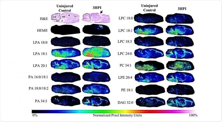 Images of sagittal brain sections of lipid species obtained by matrix-assisted laser desorption ionization imaging mass spectrometry from an uninjured control and a three hours postinjury (HPI) rat pseudocolored by intensity to demonstrate the brain-wide change in lipid levels after injury. Images of hemotoxylin and eosin (H&E) sections from adjacent sections highlight the brain regions used for mean signal intensity analysis. (Image Credit: The American Journal of Pathology)


.png)


No hay comentarios:
Publicar un comentario