Volume 23, Number 12—December 2017
Dispatch
Phylogenetic Characterization of Crimean-Congo Hemorrhagic Fever Virus, Spain
On This Page
Eva Ramírez de Arellano, Lourdes Hernández, M. José Goyanes, Marta Arsuaga, Ana Fernández Cruz, Anabel Negredo1, and María Paz Sánchez-Seco1
Abstract
Two cases of Crimean-Congo hemorrhagic fever were reported in Spain during 2016. We obtained the virus from a patient sample and characterized its full genomic sequence. Phylogenetic analysis indicated that the virus corresponds to the African genotype III, which includes viruses previously found in West and South Africa.
Crimean-Congo hemorrhagic fever (CCHF) is a severe disease transmitted to humans mainly by ticks, primarily of Hyalomma spp. This zoonotic disease is caused by CCHF virus (CCHFV), a nairovirus in the family Bunyaviridae, which was detected first in Crimea in 1944 and 25 years later in the Democratic Republic of the Congo. Since the beginning of the 21st century, CCHFV has been spreading from disease-endemic areas to new regions previously considered free of the disease, particularly in areas where Hyalomma spp. ticks are present. In nature, CCHFV usually circulates between asymptomatic animals and ticks in an enzootic cycle.
CCHFV has a negative-sense and tripartite RNA genome (small [S], medium [M], and large [L] segments) with high genetic diversity. The sequences of the S segment, which is the most conserved at the nucleotide level, could be distributed in 6 lineages (1–6). Each genetic lineage has been linked to geographic regions in Africa, the Middle East, Asia, and Europe, where Hyalomma spp. ticks are present (7). Genotypes I, II, and III have been described in Africa; genotype IV in Asia; and genotypes V and VI in Europe. However, strains have moved between geographic regions; migrating birds, unregulated wildlife trade, livestock import and export, and a global movement of humans could have dispersed the virus or CCHFV-infected ticks (8).
In western Europe, the presence of the virus had only been detected indirectly by means of serologic methods in the serum of 2 people from southern Portugal (9). However, genotype III CCHFV was detected in ticks from deer captured in western Spain in 2010 and ticks from birds migrating from Morocco in 2013 (10,11). No cases among humans in Spain had been reported until 2016, when 2 autochthonous cases were diagnosed (12). Here, we report the complete genomic sequence of the virus from 1 of these case-patients and show the phylogenetic relationships among the 3 segments.
We previously detected CCHFV in serum samples from the 2 patients in Spain with autochthonous CCHV (12); the index case-patient died. For this study, we obtained viral RNA (3.6 × 10⁷ copies/mL) from the secondary case-patient in a sample taken 4 days after onset of symptoms by using the QIAamp viral RNA Mini kit (QIAGEN, Hilden, Germany). We then amplified the virus in a single-step reverse-transcription PCR by using the SuperScript III One-Step RT-PCR system with the Platinum Taq High Fidelity DNA Polymerase kit (Invitrogen Life Technologies, Barcelona, Spain) with overlapping primers throughout the complete genome (Table). PCR conditions were amplification at 52.5°C for 30 min, 94°C for 2 min, and then 40 cycles at 94°C for 15 s, 48°C –55°C for 30 s, and 68°C for 1 min/kb, with a final extension cycle at 68°C for 5 min. We designed primers for each segment in most conserved regions after aligning available CCHFV sequences from all genotypes retrieved from GenBank (Table). We directly sequenced purified amplicons by using additional internal primers (data not shown). We assembled and analyzed the consensus sequence of each segment by using SeqMan Pro from the Lasergene Suite 12 (DNASTAR Inc., Madison, WI, USA). To get complete finished genomes, we used a hybrid-capture method as described by Blackley et al. (13) by using probes designed against the CCHFV sequences obtained from amplicon sequencing.
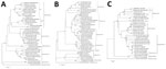
Figure. Phylogenetic analysis of Crimean-Congo hemorrhagic fever virus from patient in Spain, 2016, compared with reference sequences. A) Small segment (1,450 bp); B) medium segment (4,497 bp); C) large segment (11,829 bp)....
To characterize the complete CCHFV genome, we performed a phylogenetic analysis of the full S, M, and L segments (Figure). The 3 segments were aligned by using ClustalW in MEGA 5.2 (http://www.megasoftware.net) and representative available CCHFV sequences from GenBank of all genotypes. We generated a phylogenetic tree by using neighbor-joining algorithms and analyzed 1,000 replicates for bootstrap testing. GenBank accession numbers for sequences used in this study are MF287636 for the S fragment, MF287637 for the M fragment, and MF287638 for the L fragment.
The nucleotide sequence of the different CCHFV segments from the infected patient we analyzed in this study showed 99% identity with the Sudan AB1–2009 CCHFV strain (5) in S, M, and L segments (GenBank accession nos. HQ378179.1, HQ378187.1, and HQ378183.1, respectively) (Figure) and were grouped within genotype III (Africa 3). CCHFV found in ticks from Spain in 2010 and from birds from Morocco in 2011 also clustered in this group (10,11). In addition, no reassortant segment has been found in the analysis of the full genome, even though reassortant strains have been described in this genotype (2).
The results of the sequence analysis we describe corroborate our previous results (12), obtained by analyzing a small fragment in the S segment, showing that CCHFV from genotype III (Africa III) is circulating in southwestern Europe. CCHFV circulating in Spain caused 2 autochthonous cases that resulted in the death of the index case-patient and a serious illness in the second case-patient, providing evidence of its pathogenicity. The risk for infection in Spain is considered low, but human infection caused by the bite of an infected tick has occurred 6 years after the virus was discovered in ticks (10). Because the virus is circulating in Spain, additional studies will be required to establish the distribution of the virus in this country.
Dr. Ramirez de Arellano is a postdoctoral researcher who has worked in the Arbovirus and Imported Viral Diseases Laboratory of the National Center of Microbiology (Madrid, Spain) since 2011. Her primary research interests are detection of emerging viruses that circulate in Spain, for which she has implemented molecular biology tools.
Acknowledgments
We thank Amir Gacem for reviewing the grammar.
This study was partially funded by ISCIII, Projects RD12/0018/0006 and RD16CIII/0003/0003, “Red de Enfermedades Tropicales,” Subprogram RETICS Plan Estatal de I+D+I 2013-2016, and co-funded by FEDER “Una manera de hacer Europa.” The finished genomes were obtained with support from the US Defense Biological Product Assurance Office.
References
- Chamberlain J, Cook N, Lloyd G, Mioulet V, Tolley H, Hewson R. Co-evolutionary patterns of variation in small and large RNA segments of Crimean-Congo hemorrhagic fever virus. J Gen Virol. 2005;86:3337–41. DOIPubMed
- Deyde VM, Khristova ML, Rollin PE, Ksiazek TG, Nichol ST. Crimean-Congo hemorrhagic fever virus genomics and global diversity. J Virol. 2006;80:8834–42. DOIPubMed
- Anagnostou V, Papa A. Evolution of Crimean-Congo hemorrhagic fever virus. Infect Genet Evol. 2009;9:948–54. DOIPubMed
- Carroll SA, Bird BH, Rollin PE, Nichol ST. Ancient common ancestry of Crimean-Congo hemorrhagic fever virus. Mol Phylogenet Evol. 2010;55:1103–10. DOIPubMed
- Aradaib IE, Erickson BR, Karsany MS, Khristova ML, Elageb RM, Mohamed ME, et al. Multiple Crimean-Congo hemorrhagic fever virus strains are associated with disease outbreaks in Sudan, 2008-2009. PLoS Negl Trop Dis. 2011;5:e1159. DOIPubMed
- Singh P, Chhabra M, Sharma P, Jaiswal R, Singh G, Mittal V, et al. Molecular epidemiology of Crimean-Congo haemorrhagic fever virus in India.Epidemiol Infect. 2016;15:1–4.PubMed
- Hoogstraal H. The epidemiology of tick-borne Crimean-Congo hemorrhagic fever in Asia, Europe, and Africa. J Med Entomol. 1979;15:307–417. DOIPubMed
- Bente DA, Forrester NL, Watts DM, McAuley AJ, Whitehouse CA, Bray M. Crimean-Congo hemorrhagic fever: history, epidemiology, pathogenesis, clinical syndrome and genetic diversity. Antiviral Res. 2013;100:159–89. DOIPubMed
- Filipe AR, Calisher CH, Lazuick J. Antibodies to Congo-Crimean haemorrhagic fever, Dhori, Thogoto and Bhanja viruses in southern Portugal. Acta Virol. 1985;29:324–8.PubMed
- Estrada-Peña A, Palomar AM, Santibáñez P, Sánchez N, Habela MA, Portillo A, et al. Crimean-Congo hemorrhagic fever virus in ticks, Southwestern Europe, 2010. Emerg Infect Dis. 2012;18:179–80. DOIPubMed
- Palomar AM, Portillo A, Santibáñez P, Mazuelas D, Arizaga J, Crespo A, et al. Crimean-Congo hemorrhagic fever virus in ticks from migratory birds, Morocco. Emerg Infect Dis. 2013;19:260–3. DOIPubMed
- Negredo A, de la Calle-Prieto F, Palencia-Herrejón E, Mora-Rillo M, Astray-Mochales J, Sánchez-Seco MP, et al.; Crimean Congo Hemorrhagic Fever@Madrid Working Group. Autochthonous Crimean-Congo Hemorrhagic Fever in Spain. N Engl J Med. 2017;377:154–61. DOIPubMed
- Blackley DJ, Wiley MR, Ladner JT, Fallah M, Lo T, Gilbert ML, Gregory C, et al. Reduced evolutionary rate in reemerged Ebola virus transmission chains. Sci Adv. 2016;29;2:e1600378.
Figure
Table
Cite This Article1These authors contributed equally to this article.


.png)


No hay comentarios:
Publicar un comentario