Coronavirus: CT scans of chest could help in early diagnosis

Researchers have managed to radiologically view the novel coronavirus infected individuals, and this could be a step towards early detection of this virus and diagnosis of those infected by it. The study titled, “CT Imaging Features of 2019 Novel Coronavirus (2019-nCoV),” was published this week in the journal Radiology.
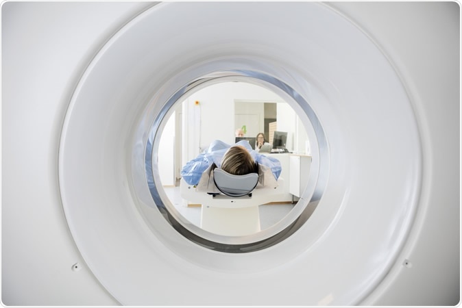
CT Scan. Image Credit: Tyler Olson / Shutterstock
The Novel coronavirus is on a rampage around the world. Its epicentre in Wuhan, (Hubei province) China is one of the worst affected regions with this deadly virus with almost ten thousand infected and nearly 500 dead from the infection.
The infection, despite high level control has managed to spread to several other nations including United Kingdom, France, Germany, Australia, Vietnam, United States, Philippines, India, Nepal and others. The World Health Organization has declared the virus to be a global threat and all steps are being taken to fight the spread of the virus. The WHO had been first notified of this infection on the 31st of December 2019.
Coronavirus infection typically manifests like viral pneumonia with fever, cough and breathlessness. The virus resembles the SARS virus (Severe Acute Respiratory Syndrome) and the MERS virus (Middle East Respiratory Syndrome).
Lead author of the study Michael Chung, assistant professor in the Department of Diagnostic, Interventional and Molecular Radiology in the Mount Sinai Health System in New York, in a statement said, “Early disease recognition is important not only for prompt implementation of treatment, but also for patient isolation and effective public health surveillance, containment and response.”
For this study the team looked at the typical chest CT scan findings of the patients infected with the 2019-nCoV in China. This was a case series with the objective of identifying the radiological features of the viral infection. For this study the team included 21 patients (13 men and 8 women) who were admitted to three hospitals in three provinces in China between 18th and 27th January 2020. The patients were aged between 29 years and 77 years and had an average age range of 51.2 years. The patients’ respiratory secretions were tested and they were all confirmed to be infected with the novel coronavirus.
CT scan images of these patients’ chests revealed certain common features including, ground glass opacities and consolidation features over the lungs. The team also noted the number of lobes of the lungs that were affected with ground glass opacities and consolidation and the degree of involvement of each of these lobes of the lungs. Based on these the team also looked at the overall total severity scores. They also noted absence of nodules in the lungs and fluid in the pleural cavity (pleural effusion). There was involvement of some of the lymph nodes of the thorax and these were abnormal in size and shape, they wrote. Some of the patients also had underlying lung diseases such as fibrosis and emphysema. These features were all recorded for the patients with the coronavirus infections. The team found some additional features such as crazy paving patterns and distribution of the changes in the peripheral parts of the lungs. There may be absence of cavity formation within the lungs along with some clear-cut nodules, they wrote. Pleural effusions and lymphadenopathy may also be absent they wrote.
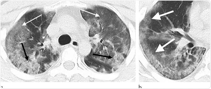
29-year old male with unknown exposure history, presenting with fever and cough, ultimately requiring intensive care unit admission. (a) Axial thin-section non-contrast CT scan shows diffuse bilateral confluent and patchy ground-glass (solid arrows) and consolidative (dashed arrows) pulmonary opacities. (b) The disease in the right middle and lower lobes has a striking peripheral distribution (arrow). Image Credit: Radiological Society of North America
Among seven of the eight patients, there was a slow progression of the lung disease with rising areas of airspace opacities, they wrote. The team wrote in their study, “Typical CT findings included bilateral pulmonary parenchymal ground-glass and consolidative pulmonary opacities, sometimes with a rounded morphology and a peripheral lung distribution.”
Dr. Chung added that not all patients showed any of these CT findings in their initial stages and thus these radiological findings may be absent in a patient. He said, “Our patient population is unique from other published series on the Wuhan coronavirus in that three of our patients had normal initial chest CTs. One of these patients progressed three days later and developed a solitary nodular ground-glass lesion in the right lower lobe, indicating this pattern may represent the very first radiologically visible manifestation of disease in some patients infected with Wuhan coronavirus.” He explained that another patient had a normal chest initially and four days after the first chest CT scan. He added, “This suggests that chest CT lacks complete sensitivity and does not have a perfect negative predictive value. We can't rely on CT alone to fully exclude presence of the virus.” He explained that the virus infection may take days to manifest as a disease and thus there may be no symptoms and no findings on CT scan in these patients.
Journal reference:
CT Imaging Features of 2019 Novel Coronavirus (2019-nCoV) Michael Chung, Adam Bernheim, Xueyan Mei, Ning Zhang, Mingqian Huang, Xianjian Zeng, Jiufa Cui, Wenjian Xu, Yang Yang, Zahi Fayad, Adam Jacobi, Kunwei Li, Shaolin Li, and Hong Shan Radiology, https://pubs.rsna.org/doi/10.1148/radiol.2020200230


.png)


No hay comentarios:
Publicar un comentario