Nanostar Applications in Biomedicine
Nanostars are a form of anisotropic, star-shaped nanoparticle, of 1-100 nm in diameter. They possess a spherical core with multiple branches of adjustable length, width and sharpness.
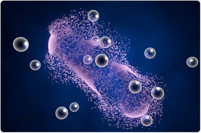
 Image Credit: Kateryna Kon / Shutterstock
Nanostars can be made from various metals, including gold, silver, platinum and palladium, among others. Nanostars may also be grown from a mixture of these and other materials such as iron oxide or silica.
The reactivity of nanoparticles that are used in catalysis, such as platinum, may be improved by shaping into a star in order to increase the surface-to-volume ratio and reactivity of the particle.
Nanostars created from plasmonic materials such as gold or silver present the most interesting applications in biology, as the surface plasmon resonance of a nanoparticle can be fine-tuned into the tissue transparency window of biological tissue. This provides applications as both diagnostic and therapeutic agents.
As with other nanoparticles, nanostars may be coated with a variety of ligands with the aim of providing protection from protein accumulation in a biological setting, providing the ability to selectively accumulate in a particular tissue or cell using targeting biomolecules such as antibodies, or as drug delivery vehicles.
Diagnostic agents
The use of gold nanostars as diagnostic tools in diverse techniques such as photoacoustic tomography, surface enhanced Raman spectroscopy, magnetic resonance imaging, positron emission tomography, and X-ray computer tomography is currently being researched.
The locally enhanced electromagnetic field at the tips of gold nanostars amplifies Raman scattering in the region, providing a sensitive method of chemical analysis that can be used to detect the presence of particular molecules in vivo or in vitro.
Photoacoustic imaging utilizes non-ionizing laser pulses to mildly heat biological tissue which then thermoelastically expands. These small expansions are detectable as ultrasonic waves that can be used to build a three-dimensional image of up to a few centimetres deep from the skin.
Gold nanostars make excellent contrast agents for this technique, as the laser in use can be fine-tuned to match the surface plasmon resonance frequency of the nanoparticle, ensuring maximum absorption and thus heating of the particle.
Since the wavelength of the surface plasmon resonance is in the tissue transparency window, the laser can influence gold nanostars deeper within the body. The entire lymphatic system of mice has been mapped in this way.
Combining gold with other materials to create nanostars allows for the enhancement of other applications. Iron oxide nanoparticles make excellent contrast agents in magnetic resonance imaging due to their superparamagnetic properties.
Taking the spherical iron oxide nanoparticle core and growing branches made from gold allows for the combination of superparamagnetic and surface plasmon resonance properties of each material, creating a combined diagnostic agent.
Therapeutic agents
Gold nanoparticles have been proposed to make excellent radiation dose enhancement agents due to their large interaction cross section and plasmonic properties.
Photons which strike electrons belonging to the nanoparticle may subsequently generate photoelectric and Compton effects, including a phenomenon known as an Auger cascade.
During Auger cascade, inner shell electrons ejected by photons may be replaced by electrons from higher energy orbitals. Upon falling to a lower energy orbital, the electron may pass the energy difference between orbitals to a third electron, causing this one to eject.
This process has been shown to repeat as many as ten times, creating a shower of low energy electrons. These electrons are capable of going on to cause destructive events within a cancer cell.
Star shaped gold nanoparticles possess a larger surface-to-volume ratio and interaction cross-section than spherical particles, and thus act more strongly as a radiation dose enhancer.
Gold nanostars can also be used as drug delivery agents, with greater specificity and retention time than free drugs alone.
By combining drug delivery and radiation dose enhancement properties multimodal anticancer agents may be developed, which allow for fewer off-target effects and thus less negative side-effects for the patient.
Drug release can be designed to be triggered upon heating, which takes place during radiation dose enhancement.
Sources:
- Plasmonic Gold Nanostars for Multi-Modality Sensing and Diagnostics.
- Multifunctional gold nanostars for molecular imaging and cancer therapy.
- Development of nanostars as a biocompatible tumor contrast agent: toward in vivo SERS imaging.
- Nanostars shine bright for you: Colloidal synthesis, properties and applications of branched metallic nanoparticles.
Further Reading
Last Updated: Oct 8, 2018


No hay comentarios:
Publicar un comentario