Difference Between Spheroplasts and Protoplasts
Protoplasts are fungal, plant or gram-positive bacterial cells without a cell wall.
Origin of Spheroplasts and Protoplasts
Spheroplasts are created from gram-negative bacteria and only part of their cell walls are removed. Gram-positive bacteria have only one cytoplasmic membrane, while gram-negative bacterium have two membranes: the cytoplasmic and the outer membrane. Therefore, following the removal of the cell wall, protoplasts have only one membrane, while spheroplasts have two membranes.
Cell Wall
The cell wall is the defensive stress-resistant layer of a cell and the cell loses this protective shield in its absence. Both spheroplasts and protoplasts adopt a spherical shape which protect against hostile environments. However, despite this structural change, these cells are very susceptible to osmotic pressures as the cell wall is responsive to environmental ionic concentration differences. Therefore, when these cells are created within the laboratory, they must be formed with isotonic solutions.
Higher concentration of the solution outside the bacterial cell would result in swelling and eventual cellular bursting. Conversely, increased concentration inside the cell would result in cellular shriveling and eventual death.
Preparation
In the laboratory, both spheroplasts and protoplasts can be formed through mechanical or enzymatic methods based on the cell type. Fungal cells can form protoplasts after chitinase treatment, while plant cells form protoplasts following pectinase, cellulase, or xylanase treatments. Both gram-positive and negative bacterium are treated with wall-digesting lysozymes or wall-inhibiting antibiotics, such as penicillin to create protoplasts and spheroplasts.
These treatments degrade or prevent the formation of the peptidoglycan links which provide mechanical strength to the cell wall. Although these cells are mainly created within the laboratory, they can also be found naturally. These bacteria are termed “L-form” and have been found in Pseudomonas, Staphylococcus, Clostridium and Bacillus.

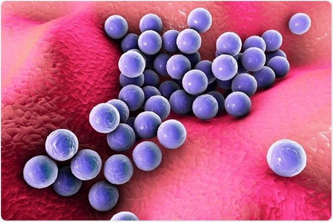
Bacteria Staphylococcus aureus on the surface of skin or mucous membrane, 3D illustration. Image Credit: Kateryna Kon / Shutterstock
Applications of Spheroplasts and Protoplasts
DNA transfection
The loss of the cell wall means that these cells can be induced to fuse with other cell types. This is useful for the transfection of DNA into animal cells. This is also useful in plant biology, allowing the fusion of protoplasts from different species, forming somatic hybrids. Plant protoplasts can also be used to recreate an entire plant from a single cell by forming a callus.
Characterising antibiotics
Spheroplasts can be used to characterise antibiotics. If a bacterium forms a spheroplast following drug treatment, then the antibiotic being tested must work through inhibiting the biosynthesis of the cellular wall. This method has led to the discovery of many antibiotics, such as cephamycin C, carbapenems and fosfomycin.
Patch clamp analysis
Giant spheroplasts (formed by prevention of cell division) can be used in patch clamp analysis, which is useful to characterise bacterial ion channels. This method monitors the current through an ion channel. While a singular bacterium is too small for this assay, giant spheroids are large enough to allow patch-clamp recording. These giant spheroplasts are grown in the presence of cephalexin, which prevents the cell from splitting and dividing, forming “snakes” with a single membrane and cytoplasm. These snakes can then have their cell walls removed and the resultant spheroid can be used in the patch-clamp analysis.
Sources
Further Reading
Last Updated: Oct 29, 2018


No hay comentarios:
Publicar un comentario