 Centers for Disease Control
and Prevention
PHIL Images From This Week
|
12/04/2015 08:00 AM EDT
This image depicts an Indonesian Field Epidemiology
Training Program (FETP) resident, as she was walking through a plantation on the way to Semono
Village, in Purworejo, Indonesia, during a malaria
outbreak investigation in December, 2014. |
|
12/03/2015 08:00 AM EDT
This image depicts an Indonesian Field Epidemiology
Training Program (FETP) resident, as she was walking through a plantation on the way to Semono Village, in Purworejo, Indonesia, during a malaria outbreak
investigation in December, 2014.
|
|
12/02/2015 08:00 AM EDT
Produced by the National Institute of Allergy and
Infectious Diseases (NIAID), this scanning electron micrograph (SEM) of a dry-fractured Vero cell revealed its contents and the ultrastructural details at the site of an opened vacuole, inside of which you can see numerousCoxiella burnetii bacteria undergoing rapid replication. |
|
12/01/2015 08:00 AM EDT
The woman pictured here was choosing an array of
healthy fruits and vegetables at a mobile produce market, and placing her choices into a yellow plastic basket she’d held in her left hand. Here she'd chosen a
bright yellow squash, which is a food that has been
shown to be high in its dietary fiber and vitamin C content. |
|
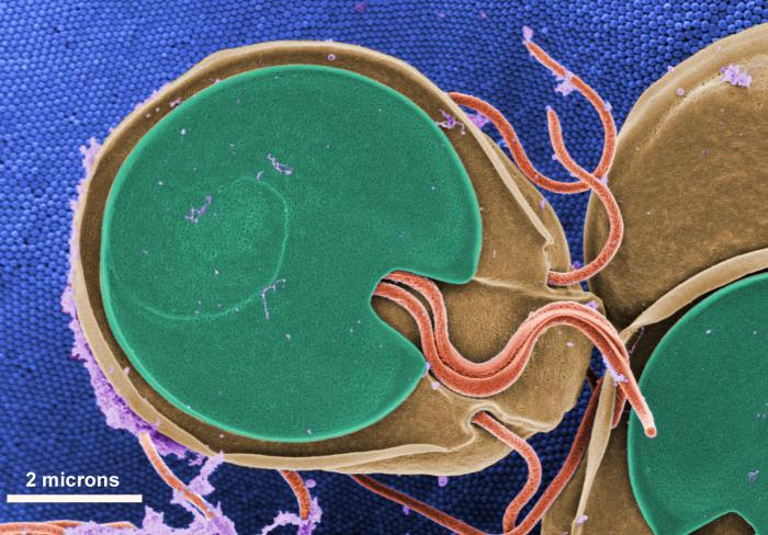
11/30/2015 08:00 AM EDT
This scanning electron micrograph (SEM) revealed
the ventral surface of a Giardia muristrophozoite that had settled atop the mucosal surface
of a rat's intestine. Note the microvilli, which can be
seen in the background,
as tiny rounded structures that are approximately
0.15 microns in diameter. |


.png)


No hay comentarios:
Publicar un comentario