jueves, 8 de noviembre de 2018

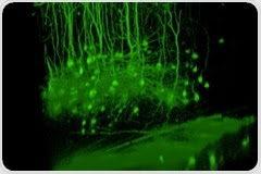
Multiphoton microscope for neurobiology applications announced by Bruker at Neuroscience 2018

Multiphoton microscope for neurobiology applications announced by Bruker at Neuroscience 2018


 November 7, 2018 
 Life Science Microscopy 
 The latest life science microscopy news from AZoNetwork 
 #ALT#Next-Generation Wide-Field, Multiphoton Microscope for Optogenetics

At the 2018 Annual Meeting of the Society for Neuroscience, Bruker announced the release of the Ultima 2Pplus next-generation multiphoton, all-optical stimulation and simultaneous imaging platform for neurobiology applications.

Read More
 
   An Introduction to Deconvolution in Optical MicroscopyAn Introduction to Deconvolution in Optical Microscopy
 
Optical microscopy is centuries old and yet remains the central technique of much of modern biological research, particularly in cell biology. It offers cell biologists the unique opportunity to examine living samples in conditions similar to those of the native state.
 
   Wide-field Fluorescence MicroscopyWide-field Fluorescence Microscopy
 
Wide-field fluorescence microscopy is a widely applied imaging technique used to examine cells and investigate their internal structures, including proteins and other cellular components.
 
 Minimizing Photobleaching in Fluorescence Microscopy
 
Minimizing Photobleaching in Fluorescence MicroscopyFluorescence microscopy is an imaging technique used to examine cells and their internal components. Specific cellular components can be labeled with fluorophores. When fluorophores absorb of a specific wavelength of light (called the excitation wavelength), their electrons move to a higher energy level.
 
 
 How do Epifluorescence Microscopes Work?
 
How do Epifluorescence Microscopes Work?Epifluorescence microscopes are a commonly used tool for studying specimens. Improved specificity and contrast provided by the application of fluorescence to the field of microscopy has stimulated the advancement of various discoveries in the biosciences. In 1843, George Gabriel Stokes described fluorescence as being characterized by the wavelength of an emitted light that is longer than the wavelength of the exciting light.
 
 
 Photobleaching in Fluorescence Microscopy
 
Photobleaching in Fluorescence MicroscopyPhotobleaching is the phenomenon when a fluorophore loses its fluorescence due to damage induced by light. This leads to loss of fluorescence and signal while imaging a sample. When light of appropriate wavelength is directed on a fluorophore, it transitions from ground state to excited singlet and triplet stages. In its excited state, it may interact with other molecules and undergo permanent covalent modifications.
 
 
 Digital Imaging in Optical Microscopy
 
Digital Imaging in Optical MicroscopySince the first prototype of the modern microscope was developed at the end of the 16th century, an immense amount of progress in the field of microscopy has allowed this tool to play a crucial role in science. For example, the modern field of digital pathology utilizes the power of microscopy to perform clinical screening and diagnostic procedures through cellular-, molecular- and genetic-imaging.
 

No hay comentarios:

Publicar un comentario