|
| | November 9, 2018 | |
| | |
| | The latest fluorescence news from AZoNetwork | |
|
|
 |
| | 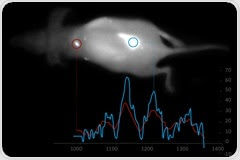 Hyperspectral Infrared Imaging Opens a New Window on Living Bodies Hyperspectral Infrared Imaging Opens a New Window on Living Bodies
Using the first (NIR) and second biological window (NIR-II) from 900-1620 nm, Photon etc.’s IR VIVO™ preclinical imager provides outstanding hyperspectral infrared imaging capabilities. It combines micron-scale spatial resolution, real-time imaging and full spectral coverage throughout small animals. Emission of several fluorophores can be isolated with a high-efficiency tunable filter and ultra-low noise scientific-grade InGaAs camera.
| |
|
|
|
|
|
| | 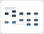 Immunofluorescence is based on the use of antibodies with fluorescent labels to bind to and thereby enable the detection of specific target antigens. Immunofluorescence is based on the use of antibodies with fluorescent labels to bind to and thereby enable the detection of specific target antigens. | |
|
| | 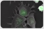 The 3D Cell Explorer-fluo can make movies involving fluorescence as well as 3D refractive index imaging to produce time-lapse images acquired from mouse embryonic stem cells. The 3D Cell Explorer-fluo can make movies involving fluorescence as well as 3D refractive index imaging to produce time-lapse images acquired from mouse embryonic stem cells. | |
|
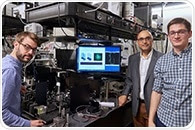
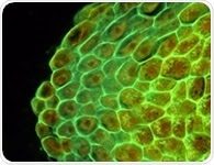
| |  Wide-field fluorescence microscopy is a widely applied imaging technique used to examine cells and investigate their internal structures. Wide-field fluorescence microscopy is a widely applied imaging technique used to examine cells and investigate their internal structures. | |
|
| |  Photobleaching is the phenomenon when a fluorophore loses its fluorescence due to damage induced by light. This leads to loss of fluorescence and signal while imaging a sample. Photobleaching is the phenomenon when a fluorophore loses its fluorescence due to damage induced by light. This leads to loss of fluorescence and signal while imaging a sample. | |
|
|
|
|


.png)


No hay comentarios:
Publicar un comentario