|
| | October 24, 2018 | |
| | |
| | The latest life science microscopy news from AZoNetwork | |
|
|
 |
| |  Label-Free Long-Term 3D Imaging of Mitochondria Label-Free Long-Term 3D Imaging of Mitochondria
Mitochondria are one of the most important cell organelles. They are organized in extremely dynamic networks and, in addition to supplying the cell with energy, they have an important role in apoptosis, metabolism, and degenerative diseases. Until now it was believed that, unless specifically stained, they were not visible in microscopy.
| |
|
|
|
|
|
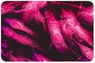
| |  Fluorescence microscopy is an imaging technique used to examine cells and their internal components. Specific cellular components can be labeled with fluorophores. Fluorescence microscopy is an imaging technique used to examine cells and their internal components. Specific cellular components can be labeled with fluorophores. | |
|
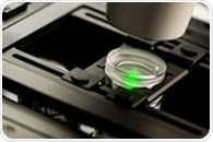
| |  A novel optical microscope utilizing diffraction tomography to generate 3D holographic images of unlabelled live cells is now available from Tomocube. Called HT-1, the new microscope delivers quantitative nanoscale, real-time, dynamic images of individual living cells quickly and simply without any sample preparation. A novel optical microscope utilizing diffraction tomography to generate 3D holographic images of unlabelled live cells is now available from Tomocube. Called HT-1, the new microscope delivers quantitative nanoscale, real-time, dynamic images of individual living cells quickly and simply without any sample preparation. | |
|
| |  Holotomography uses lasers to measure the refractive index in all three dimensions. As the refractive index of different objects inside a cell varies, this method can be used as a contrast agent to visualise different components inside a cell. Holotomography uses lasers to measure the refractive index in all three dimensions. As the refractive index of different objects inside a cell varies, this method can be used as a contrast agent to visualise different components inside a cell. | |
|
| |  In a revolutionary study, an LMU research team headed by Ralf Jungmann has shown that the application of chemically-modified DNA aptamers as protein markers enables one to improve the power of super-resolution fluorescence microscopy as an imaging instrument. In a revolutionary study, an LMU research team headed by Ralf Jungmann has shown that the application of chemically-modified DNA aptamers as protein markers enables one to improve the power of super-resolution fluorescence microscopy as an imaging instrument. | |
|
|
|
|


.png)


No hay comentarios:
Publicar un comentario