
Cool Images: An Independence Day-Inspired Collection

In case you missed the fireworks this weekend, we’ve put together a collection of firework-like images from basic research studies.
Viral Electricity
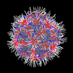
This patriotic Koosh ball is an adeno-associated virus. Most people will come into contact with the virus at some point in their lives, and they’ll probably never know it. Even though it doesn’t cause disease—in fact, because it doesn’t cause disease—this virus is scientifically important. Researchers hope to harness the virus’ ability to enter cells and hijack genes and to use it to to deliver gene therapy. This image, created with the software DelPhi, shows which parts of the virus are positively charged (blue) and which parts are negatively charged (red). The charge of a molecule—like the charge of this virus—influences the way it behaves. In addition to helping researchers understand how viruses might enter cells, images like this one could help them understand how molecules interact with each other as well as drugs.
Dynamic Dendrites
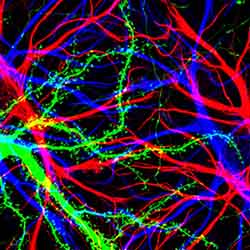
These fireworks aren’t in the sky—they’re in your brain. The lightning-like lines are cells in the hippocampus, the small, crescent-shaped part of the brain involved in memory and movement. The dotted green lines are dendrites, the branched ends of neurons that receive signals from other electricity-transmitting nerve cells and carry them through the body. Glial cells, which provide support and nutrition to neurons, are shown in red and blue.
The Genetic Network
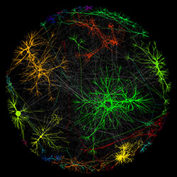
This collection of multicolored starbursts might remind you of a static electricity demo in a science museum, but it’s actually a digital model of genes interacting in networks. Created using the software Cytoscape, this image shows some of the genetic interactions in a yeast cell. Each dot represents a gene, and each line indicates a connection between genes. Just as people with similar interests stay in contact through social networks, the color-coded groups of genes accomplish specific goals—say, responding to a recent meal, fighting off infection or helping the cell divide. Scientists map gene networks to study, for example, different kinds of diseases and cells’ responses to changes in their environment.
Fluorescent Fireworks
NIH Director Francis Collins calls this video “fireworks under a microscope.” The astounding display shows microtubules assembling and branching during cell division. Microtubules are the thin, hollow rods that act as the supportive skeleton of cells. Before cells can divide, these tiny tubes must organize and grow. As the microtubules grow, they glow red. As new ends branch off, they shine bright green. The researchers who captured this video showed for the first time that not only do new microtubules branch during cell division, but they do so very rapidly, going from a few branches to hundreds in a matter of minutes
Living Liver
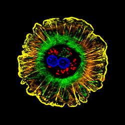
This tiny explosion is a digital micrograph of a hepatocyte, the type of cell that makes up around 80 percent of your liver. Hepatocytes break down fats, proteins and sugars, and they remove toxins from the body. Detoxifying is difficult work and can sometimes damage the liver. But, remarkably, hepatocytes have the ability to quickly regrow after injury. Researchers are investigating whether the liver’s self-repair potion—hepatocyte growth factor—can be used to heal damage to other organs in the body.
Electrostatic image: Emil Alexov, Clemson University; hippocampus image: Barbara Calabrese and Shelley Halpain, University of California, San Diego; Cytoscape image: Keiichiro Ono and Trey Ideker, University of California, San Diego; microtubule video: Sabine Petry, Princeton University; hepatocyte image: Donna Beer Stolz, University of Pittsburgh.


.png)


.png)


No hay comentarios:
Publicar un comentario