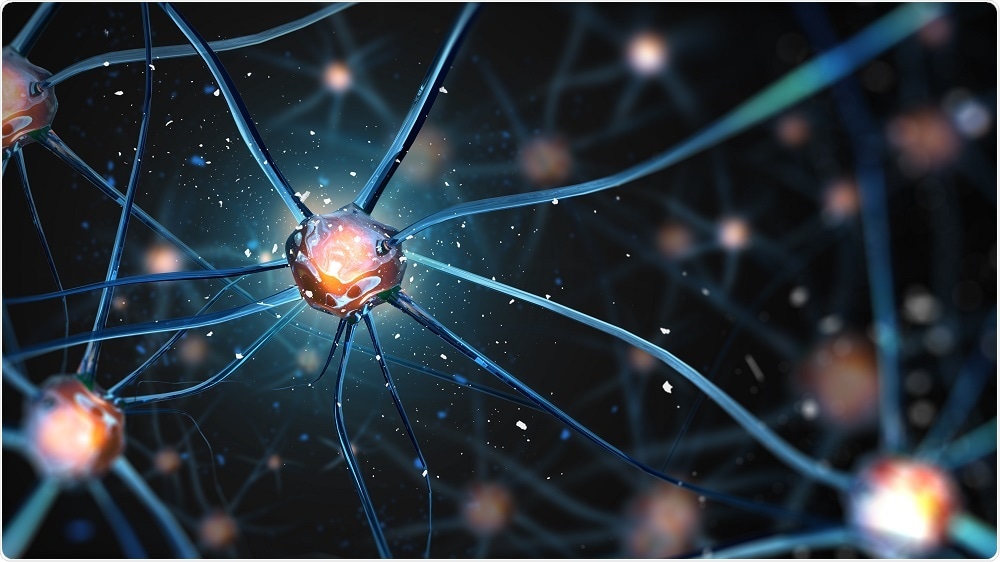
Scientists discover abnormal brain networks in children with autism
A new study has shown that pre-school children with autism spectrum disorder (ASD) have abnormal connections between certain brain networks that can be visualized using an MRI technique called diffusion tensor imaging (DTI).
 Credit: Andrii Vodolazhskyi/Shutterstock.com
Brain networks are areas of the brain connected by white matter tracts that interact to carry out different functions and the DTI technique provides important information about the state of the brain’s white matter.
The study authors say the findings could help lead to the development of treatments for ASD. Early intervention and diagnosis of ASD is key, since younger patients usually benefit the most from treatment.
As reported in the journal Radiology, the team compared DTI results obtained from 21 pre-school children (aged a mean of 4.5 years) with those of 21 children of a similar age who did not have ASD.
The results showed that the children with ASD had significant differences in components of the basal ganglia network, compared with the children who did not have ASD.
The basal ganglia network is a system that plays an important role in behavior. In addition, between-group differences were seen in the paralimbic-limbic network, which is also involved in regulating behaviour.
The results suggest that the different patterns may underlie the abnormal brain development seen in pre-schoolers with ASD and may play a role in the mechanisms involved in the disorder. The discovery may also guide the development of potential imaging biomarkers for pre-schoolers with ASD.
"The imaging finding of those 'targets' may be a clue for future diagnosis and even for therapeutic intervention in preschool children with ASD," says Ma.


.png)


No hay comentarios:
Publicar un comentario