Insulin Signaling Pathway

Insulin signaling is the pathway that regulates glucose homeostasis through the control of important processes such as glucose and lipid metabolism.
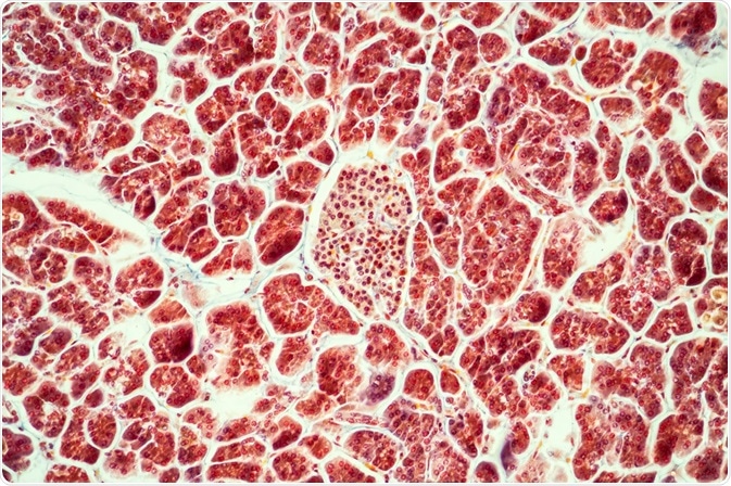 Credit: Dr. Norbert Lange/Shutterstock.com
When insulin binds to an insulin receptor (IR) in mammalian cells, a wide range of complex biological effects are seen. The IR β subunit tyrosine kinase is activated, which then phosphorylates IR substrate (IRS) proteins.
IRS-1 and IRS-2 are the two primary substrates that undergo phosphorylation and these activate the phosphatidylinositol 3-kinase (PI3K)-Akt pathway, which plays a major role in the actions of insulin, mainly through activation of the Akt/PKB and the PKC cascades. IRS-1 and IRS-2 also activate the Ras-mitogen-activated protein kinase (MAPK) pathway, which works in conjunction with the PI3K pathway to regulate cell proliferation.
Phosphorylation cascade
At least six substrate proteins are known to be phosphorylated by IR. All have the ability to interact with five main forms of the P13K regulatory subunit. These subunits interact with three types of PI3K catalytic subunit. This leads to the production of phosphatidylinositol-3,4,5-triphosphate (PIP3), which, via PDK1 kinase, induces activation of the three known isoforms of Akt.
These different combinations of associations enable immense variations in and tailoring of insulin signaling in both health and disease states.
Physiological effects of insulin signaling
When insulin signals to Akt, various metabolic effects are seen. In muscle cells and fat cells, a number of kinases phosphorylate the Rab-GTPase-activating proteins AS160 (TBC1D4) and TBC1D1, which induces the uptake of glucose.
In liver cells, phosphorylation and inactivation of the transcription factor Fox01 by Akt is at least partly responsible for the repression of genes required for glucose production. Lipid production is also increased through kinase-dependent pathways that involve SREBP1c, PI3K, PDK1 and PKCλ/ζ.
Additionally, glycogen synthesis is increased through the GSK3β/glycogen synthase pathway and lipolysis is inhibited through the Akt/PDE3B pathway. Insulin signaling has also been shown to induce cell proliferation and to prevent cell death. Akt and a number of other kinases phosphorylate the tuberous sclerosis complex, which suppresses its ability to activate GTPase. This causes activation of the mTORC1 activator Rheb, which leads to increased cell proliferation and induces translation. Cell proliferation is also promoted via activation of the MAP kinase cascade, which is triggered by Son-of-Sevenless (SOS). Cell death is inhibited through the Akt/BAD axis.
Regulation of insulin signaling
Multiple levels of regulatory controls influence insulin signaling and the pathways downstream of it. Feedback loops and the responses of pathways to various stimuli are amongst these mechanisms. IR activity is regulated by feedback loops. IR is dephosphorylated and its activity limited by a phosphotyrosine phosphatase called PTP1B. However, H2O2 is generated through IR signaling, which leads to inhibition of PTP1B and extended insulin signaling.
An adapter protein called Grb10 inhibits the activity of IR kinase and the phosphorylation of Grb10 by mTORC1 has been shown to enhance the inhibition of IR.IR recognition of IRS proteins is inhibited by a number of serine/threonine kinases. Serine residues of IRS-1 are phosphorylated by S6K1 and mTORC1, which prevents tyrosine being phosphorylated.
Tribbles homolog 3 associates with Akt, which inhibits its ability to be recognized by mTORC2 and PDK1. The activity of Akt is also decreased by protein phosphatase 2A. Fox01 induces the transcription of IRS-2, which is quickly reduced as a result of Akt promoting the exclusion of Fox01 from the nucleus.
On binding to their receptors, proinflammatory cytokines activate SOCS-3, which interacts with IR to prevent its recognition of IRS. Multiple stimuli induce JNK1 including cytokines and oxidative stress. Activated JNK1 phosphorylates a domain on IRS-1 which decreases the phosphorylation of tyrosine. A build up of lipids activates PKCθ, which phosphorylates IRS-1.
As well as binding to IR, SOC-3 also interacts with IRS proteins to stimulate their breakdown and decrease their signaling.
Reviewed by Liji Thomas, MD.
Sources:
Further Reading
Last Updated: Mar 5, 2018


.png)

.png)


No hay comentarios:
Publicar un comentario