
Purification, Tracking, and Characterization of Microvesicles
Introduction
Exosomes are microvesicles that are formed inside the cell. These microvesicles contain and protect a variety of protein and RNA cargos which are secreted into the bodily fluids, including blood.
Nearby and distant cells take up these exosomes where their RNA and protein cargos can modulate recipient cell activity. As a result, there has a been a great deal of interest in the ability of exosomes as vectors for gene therapy as well as diagnostics which can detect diseases on the basis of the analysis of exosomes recovered from various body fluids such as saliva, urine, and blood.
A range of useful, cost-effective products are available from Cell Guidance Systems to support your exosome research.
Exo-spin™ purification
Exosome precipitation solutions leave a significant amount of non-exosomal proteins in samples. Exo-spin™ kits use size exclusion technology, either alone or in combination with a proprietary precipitation reagent, to yield pure exosomes.
Exo-spin™ kits are available for a wide range of biological fluids, including saliva, urine, cell culture media, and blood plasma/sera.
Kits are available in midi drip-column and mini-spin column sizes and for 24 and 48 samples. Exo-spin™ kits provide outstanding quality and value-for-money.
Choosing Exo-spin™ products
The last step of any Exo-spin™ protocol utilizes size exclusion chromatography (SEC), using a proprietary resin which is calibrated to purify particles in the 30-400 nm range.
All commonly established definitions of exosome size are covered by this range - the same size range purified with ultracentrifugation, meaning that samples prepared with Exo-spin are comparable with samples obtained by ultracentrifugation..
Exo-spin™ is available in a range of configurations to fit your particular applicaiton. In the standard procedure, precipitation is followed by additional purification by SEC columns. In cases where there are high concentrations of exosomes, the precipitation step can be skipped.
Eliminating the precipitation step is indeed necessary for mass spectrometry and other similar applications.
For blood samples, the concentrations of exosome are normally of the order of 1x10¹²particles per ml, and hence there is no need for precipitation. Using product EX04 (for samples up to 1 ml) and product EX03 (for samples up to 0.1 ml), exosomes from blood sera can be directly and rapidly purified by ExoSpin™ SEC columns without the need for any precipitation.
Normally, cell culture produces yields of 1x10⁹ particles per ml or less. Integra CELLine flask is a cell culture device that can be used to raise the concentration of exosomes in the media by as much as 10-fold, preventing the need for precipitation.
When there is a need to process larger sample volumes, ExoSpin™ precipitation buffer is used to concentrate exosomes before SEC column purification. ExoSpin™ kit EX01 can be used for urine, cell culture, saliva and other low exosome concentration biological samples, and kit EX02 may be used for for blood. Regardless of the sample and the application, there is an Exo-spin™ product to meet user’s needs.
Exosome detection
A range of kits and products, to assay, track and detect microvesicles, are offered by Cell Guidance Systems.
TRIFic™ detection assay
While the TRIFic™ exosome assay is similar to an ELISA assay, there are a few key differences. For instance, there is no enzymatic reaction unlike an ELISA assay. Instead a Europium label is used to directly detect the target. In addition, the binding and detection antibody in the TRIFic™ assay are the same.
TRIFic™ exosome assays rapidly provide quantitative data from unpurified or purified samples, including direct measurement of exosomes in plasma in an easy 96-well format. These assays are now available for commonly used exosome markers – the tetraspanin proteins CD9, CD81 and CD63. Clear and consistent data is delivered by TRIFic™ exosome assays.
Attributes
The same antibody is used in the TRIFic™ exosome assay for binding the target to the assay plate as well as for detection. The assay comprises of a monoclonal antibody (labelled with biotin) attached to a streptavidin coated plate capable of capturing protein present on the exosome surface, as shown in Figure 1.
Consequently, detection is performed using an identical monoclonal antibody (labelled with Europium). Since the capture and detection antibody are identical, they need two linked copies of the same epitope to detect a signal. A perfect structure to enable signal is provided by exosomes: These connect two copies of a molecule and permit their detection in this assay.
Take, for example, CD9. Exosomes typically have several copies of CD9 that face towards the attachment surface on the assay plate and extra CD9 molecules, on the opposite side of the exosome available for detection. In contrast, a signal may not be generated by any non-specific binding of capture and detection antibodies.
The use of a Europium fluorophore offers high levels of sensitivity for the assay, which is capable of detecting slight changes in the abundance of the target CD9 protein even within complex, unpurified biological samples, such as cerebrospinal fluid and blood plasma.
- Streptavidin-coated assay plates are bound with biotinylated antibody.
- Biological samples are added. The antibody captures exosomes and any free antigen.
- Antibody labeled with Europium is added and binds particularly to exosome antigen. The epitopes of bound monomers are by now occupied and remains undetected. Reading of samples takes place on a time-resolved fluorescence plate reader.
Advantages
- Europium offers a high degree of sensitivity and hence used for detection.
- The assay detects only antigens displayed in several copies on a support within the sample (as with tetraspanins on exosomes), thus leading to increased specificity.
- Thanks to the simplicity of the assay, a high degree of reproducibility is achieved
Product data
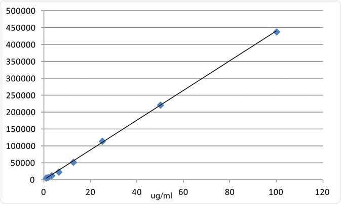
Titration of purified exosomes using TRIFic™ exosome assay shows a linear relationship between quantity of exosomes and TRF units over a wide range. The assay is able to detect sub-microgram levels of exosomes.
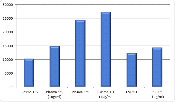
Biological samples (diluted 1:1 or 1:5 with PBS) spiked with purified exosomes demonstrates the sensitivity of the assay even in complex biological fluids such as plasma and cerebrospinal fluid.
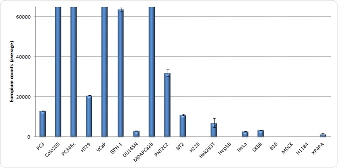
CD9 TRIFic™ exosome assay performed for a variety of cell lines. Over 80 samples plus standards can be tested with a single kit.
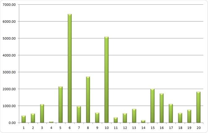
TRIFic™ exosome assay analysis of CD9 in 20 cancer patient urine samples demonstrating a wide range of exosomal content.
ExoFLARE™ tracking assay
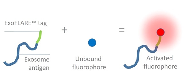
A combination of Fluorescence Activating Response Element (FLARE) protein tag and a pro-fluorophore dye is used by ExoFLARE™. The dye and protein do not fluoresce in isolation. After the protein attaches to the dye, it induces structural changes resulting in fluorescence.
An unstable bond is formed by the protein and dye with constant turnover of the dye. This leads to continuous fluorescence without the levels of photo-bleaching that is common with fluorescent proteins. This makes it possible to monitor ExoFLAREs™ for long periods of time and thus track the dye movement.
Attributes
- As mentioned before, the ExoFLARE™ system includes two components, which collectively provide unprecedented signal durability and specificity
- All ExoFLARE™ mammalian expression constructs are ready to transfect into cells. Currently, CD9, CD63 and CD81 are available
- An assay is typically carried out in a single well of a 96 well plate, and ExoFLARE™ fluorophores are provided in 100 or 1000 assays
Advantages
- Non-toxic
- Considerably brighter than fluorescent proteins
- Low background levels
- Long duration of signal without noticeable photobleaching
Use ExoFLARE™ to:
- Image individual exosomes
- Detect uptake of exosomes in non-transfected recipient cells
- Monitor long-term movement of exosomes
Kit contents
Each kit contains:
- EF Red s cell permeable dye for 100 assays
- 20 µg of a mammalian expression construct (for a single CD antigen)
- PBS buffer
- Solvent A for resuspension of dye
- Comprehensive User Guide
Product data
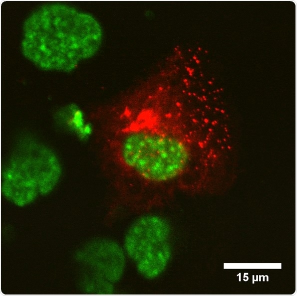
CD9
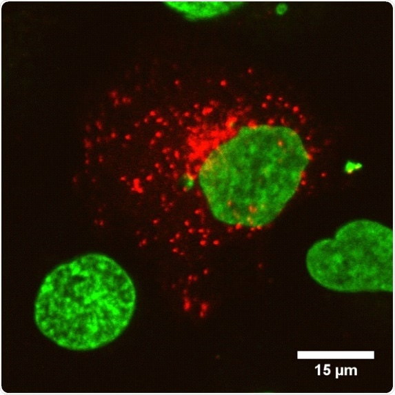
CD63
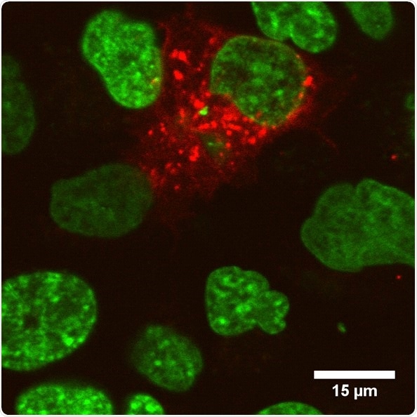
CD81
ExoFLARE™ constructs were transiently transfected into DU154N cells. ExoFLARE™ cell permeable dye was added to the media and cells were imaged using a confocal fluorescence microscope. Red = ExoFLARE™; Green = Hoechst.
Validated antibodies
Cell Guidance Systems offers exosome antigen antibodies that detect tetraspanins including the most extensively studied exosome antigens. These antibodies have been verified for exosome detection. Cell Guidance Systems supplies the same antibodies that are used in its TRIFic™ assay kit.
NTA size profiling service
To analyze microvesicles isolated from a biological sample, many different parameters are measured to build up a comprehensive characterization. The size and number distribution of exosome particles is one of the most important metrics and is commonly determined in exosome research.
A measure of sample purity can be extrapolated by integrating this measurement with the sample protein content measured e.g. by a Bradford assay.
Tracking of Brownian motion, called nanoparticle tracking analysis (NTA), is the most extensively used and accepted method for exosome size analysis. These kinds of measurements were pioneered in the field of exosomes and microvesicles using Nanosight instruments.
ZetaView® is a second-generation particle size instrument from Particle Metrix, and is used by Cell Guidance Systems exosome analysis service. Thanks to its ease of use, the instrument offers higher throughput and greater economy, enabling users to deliver an exosome analysis service that is highly cost-effective.
About the ZetaView®
ZetaView® is a widely used, high performance integrated instrument that utilizes measurements of Brownian motion, classical micro-electrophoresis, and individual exosome particle tracking.
Highly resolved size histograms and zeta potential are rapidly derived from particles and it is possible to determine concentrations of even small numbers of particles. The ZetaView®offers analysis certainty, resulting in an improved level of exosome characterization and enabling innovative discoveries in the field of exosome and microvesicle research.
About Cell Guidance Systems
Cell Guidance Systems is based in Cambridge, UK. They were founded in 2010 and opened our US office in 2011. Over the years, they have been quietly building a reputation for quality, service and innovation.
Cellgs are part of one of the most exciting biotech regions in the world. They work to bring innovations from research groups around the world (including Japan, USA, Netherlands, Italy, Singapore and the UK) to develop products that expand the possibilities of life science research.
Many of their products are truly groundbreaking: For example, ETS-embryo medium (developed in the lab of Prof Magdalena Zernicka-Goetz at Cambridge University) enables the production of artificial embryos from stem cells. PODS™ (developed in the lab of Prof Hajime Mori at Kyoto Institute of Technology) is overcoming stability limitations of proteins, offering hope for new therapies.
In addition to providing reagents and research services, they are involved in several areas of therapeutic research based on the PODS™ technology. Cell Guidance Systems are collaborating with researchers and companies in the UK, Italy and Japan to develop vaccines and therapeutics for a variety of diseases.
The pursuit of quality drives everything Cellgs do, from the components they use, the manufacturing process and the final preparation and packaging of the product. They employ rigorous quality control to ensure products function effectively.
Cell Guidance systems here to help you on your quest to discover the unknown, understand the complex, and heal the many diseases and conditions that affect us all.
Sponsored Content Policy: News-Medical.net publishes articles and related content that may be derived from sources where we have existing commercial relationships, provided such content adds value to the core editorial ethos of News-Medical.Net which is to educate and inform site visitors interested in medical research, science, medical devices and treatments.
Last updated: Oct 3, 2017 at 11:41 AM


.png)


No hay comentarios:
Publicar un comentario