SPECT PET Imaging Technology Drives Initiatives in Targeted Cancer Therapy
According to the American Cancer Society, approximately 1,688,780 new cancer cases will occur in 2017,1 half of which will result in subsequent mortality. Targeted therapeutic cancer approaches, including radiation and protein inhibitors, are common treatment options for improving the likelihood of survival.2,3 The use of selective kinase inhibitors, for example, may provide improved patient outcomes in clinical oncology.3 Imaging modalities are one of the prime methods for gaining first-hand insight into the effect of targeted treatment.
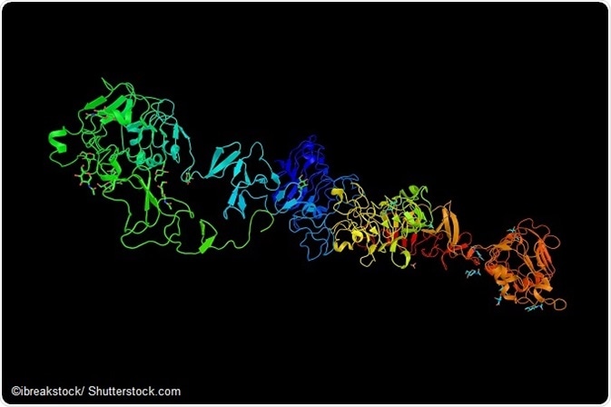
Image: Receptor tyrosine-protein kinase erbB-3 (HER3).
HER3 Overexpression: Imaging and Targeted Therapy
Human epidermal growth factor receptor 3 (HER3) is a membrane-bound protein found in the ERBB3 gene. The receptor is involved in proliferation, motility, and apoptosis suppression. In some cancers, the HER3 protein is overexpressed and can lead to disease progression and reduced effectiveness of therapy.4 Imaging of HER3 in vivo can help determine if the protein is being overexpressed and, if so, whether or not the patient would benefit from anti-HER3 immunotherapy. Blocking HER3 may provide some assistance in reducing resistance to therapy, according to some research.5
Assessment of biomarkers for evaluating the status of HER3 expression remain little explored, presenting a roadblock for selecting patients for appropriate therapies. Molecular imaging of HER3 receptors to determine overexpression may assist clinicians in evaluating a patient’s risk for resistance to therapy while enabling effective monitoring for tumor response to chemotherapy intervention.
Imaging HER3 has been a clinical challenge due to its somewhat low receptor expression in tumors (<50000 receptors/cell).6 PET provides high resolution, quantification accuracy, and high sensitivity. Image enhancement can occur when combined with single photon emission computed tomography (SPECT) imaging.
Affibody molecules, when used during PET SPECT imaging, can also offer high imaging contrast. Previous research found that positron-emitting radionuclide gallium-68 featuring a half-life of 67.8 minutes may be appropriate for HER3 imaging. PET, according to the authors, may provide high accuracy for both molecular diagnostics and quantitative information of target expression.4
Recent Research
According to Pieve et al. in the journal Bioconjugate Chemistry, the development of high target affinity positron emission tomography (PET) probes is necessary for imaging HER3 in vivo, which can help stratify patients at risk for therapeutic resistance.7
A study reported in the same paper looked at two strategies for radiolabeling the HER3 affibody molecule for targeting. Specifically, Pieve et al. examined affibody-based agents for 18 F PET imaging of HER3 expression.
One strategy used Bruker’s Albira Si, a PET SPECT molecular imaging system.8 SPECT imaging has been used in previous research to evaluate everything from novel sentinel lymph node imaging agents9 to studying targeted radiotherapy.10 A combined imaging modality with CT imaging can also offer anatomical landmarking as well as helping researchers assess changes in tumor progression.11
Researchers used female NCr athymic mice for in vivo testing, injecting each animal with MCF-7 or MDA-MB-231 cells in 30% Matrigel. Estrogen pellets (17β-estradiol) were inserted into MCF-7 cells prior to inoculation. Researchers allowed tumors to grow for approximately 3 weeks. Following growth, researchers utilized the Albira Si imaging system to examine changes at the molecular level.
After 1 hour of radioconjugate injection, a whole PET static imaging was performed for 10 minutes. Imaging data was corrected for dead-time count losses, PET nonuniformity, and physical decay. Using an X-ray tube setup at 45 kV, whole body standard high resolution CT scans were performed. Evaluation of the final images was assisted by PMOD software. The entire Bruker package was able to provide this in-depth analysis efficiently.
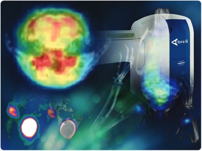
Bruker’s Albira Si: First Commercial SiPM-based PET
The imaging of HER3 in this trial was essential for assessing a therapeutic target in cancer. Utilization of such an approach in clinical oncology may provide effective risk stratification for patients undergoing therapy, particularly in regard to the patient’s individual response to treatment.
In this study, researchers used Albira Si, a PET/SPECT/CT imaging system offered by Bruker. For in vivo studies that require thorough understanding of underlying biological processes as well as the progression of disease, a system like the Albira Si can be an invaluable tool, especially in trials with mice and other small animals.
Bruker’s Albira Si is the first commercial SiPM-based PET with integrated SPECT and CT. Additionally, this system is compatible with magnetic resonance imaging, a capability that can only enhance imaging results in preclinical and clinical trials.
References:
- Siegel RL, Miller KD, Jemal A. Cancer Statistics, 2017. CA Cancer J Clin. 2017 Jan;67(1):7-30.
- ASTRO. Advances in radiation therapy have improved survival rates for early stage lung cancer patients. https://www.astro.org/News-and-Publications/News-and-Media-Center/News-Releases/2016/Advances-in-radiation-therapy-have-improved-survival-rates-for-early-stage-lung-cancer-patients/. Published September 25, 2016.
- Gross S, Rahal R, Stransky N, Lengauer C, Hoeflich KP. Targeting cancer with kinase inhibitors. J Clin Invest. 2015;125(5):1780-1789.
- Rosestedt M, Andersson KG, Mitran B, et al. Affibody-mediated PET imaging of HER3 expression in malignant tumours. Sci Rep. 2015;5:15226.
- Li C, Brand TM, Iida M, et al. Human epidermal growth factor receptor 3 (HER3) blockade with U3-1287/AMG888 enhances the efficacy of radiation therapy in lung and head and neck carcinoma. Discov Med. 2013;16(87):79-92.
- Robinson MK, Hodge KM, Horak E, et al. Targeting ErbB2 and ErbB3 with a bispecific single-chain Fv enhances targeting selectivity and induces a therapeutic effect in vitro. Br J Cancer. 2008;99(9):1415-1425.
- Pieve CD, Allott L, Martins CD, et al. Efficient [18F]AlF Radiolabeling of ZHER3:8698Affibody Molecule for Imaging of HER3 Positive Tumors. Bioconjugate Chemistry. 2016;27(8):1839–1849.
- AlbiraSi. Bruker. https://www.bruker.com/products/preclinical-imaging/pet-spect-ct/albirasi/overview.html.
- Ocampo-García B1, Ferro-Flores G, Morales-Avila E, Ramírez Fde M. Kit for preparation of multimeric receptor-specific ⁹⁹mTc-radiopharmaceuticals based on gold nanoparticles. Nucl Med Commun. 2011;32(11):1095-1104.
- Vilchis-Juárez A, Ferro-Flores G, Santos-Cuevas C, et al. Molecular targeting radiotherapy with cyclo-RGDFK(C) peptides conjugated to 177Lu-labeled gold nanoparticles in tumor-bearing mice. J Biomed Nanotechnol. 2014;10(3):393-404.
- Davison CA, Chapman SE, Sasser TA, et al. Multimodal optical, X-ray CT, and SPECT imaging of a mouse model of breast cancer lung metastasis. Curr Mol Med. 2013;13(3):368-376.
About Bruker
Bruker delivers the world's most comprehensive range of research tools enabling life science, materials science, analytical chemistry, process control and clinical research. Bruker is also the leading superconductor magnet and ultra high field magnet manufacturer for NMR and MRI solutions.
Sponsored Content Policy: News-Medical.net publishes articles and related content that may be derived from sources where we have existing commercial relationships, provided such content adds value to the core editorial ethos of News-Medical.Net which is to educate and inform site visitors interested in medical research, science, medical devices and treatments.
Last updated: Jul 27, 2017 at 5:50 AM


.png)


No hay comentarios:
Publicar un comentario