Advantages and Applications of Bruker’s MRI CryoProbe Series
Introduction
The MRI CryoProbe series from Bruker has superior resolution and provides the ultimate image with unmatched performance. MRI CryoProbes are equipped with very low temperatures, closed-cycle cooled RF-coils and preamplifiers, delivering an increased signal-to-noise ratio (SNR) by a factor of 2.5 in routine in vivo MRI applications at 9.4 T when compared to standard room temperature 1H RF-coils.
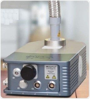
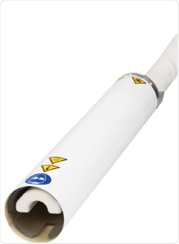
MRI Cyroprobe.
Pioneering performance
Bruker is recognized as the principal innovator in magnetic resonance and originally launched the CryoProbe technology more than a decade ago. Today, the company has installed over 1200 CryoProbes all across the globe, providing unrivaled performance by reliably achieving increased SNR. This increased sensitivity results in shorter measurement times and improved throughput with reduced animal stress.
Unique technology for excellent results
Cryogenically cooled RF-coils and preamplifiers are used in Bruker’s MRl CryoProbe technology. A closed-cycle refrigeration system is used to cool these RF-coils and preamplifiers, thereby improving the coil performance and significantly reducing the noise contribution of the associated electronics.
The MRl CryoProbe handles and supervises animals in a very similar manner to that of standard room temperature RF-coils. Cooling the MRI CryoProbe can be achieved outside the magnet, allowing the scanner time to be used optimally.
MRI CryoCoils are advantageous in many ways over standard room-temperature RF-coils:
- Higher resolution in vivo up to 20 µm
- Increase in SNR by up to a factor of 5
- Shorter durations of anesthesia
- Shorter measurement times
- Shorter measurements directly enable higher productivity and lower costs per sample
- New applications cannot be accessed with room temperature RF-coils (high resolution, fMRI, etc.)
Applications
Dynamic 13C-MRS of the mouse brain
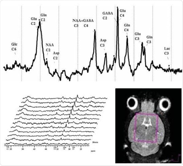
Localized 13C(1H} spectroscopy (ISIS) over time of mouse brain after orally administrated [1-13Cl] glucose. Courtesy: H. Terasawa, Kumamoto University, Kumamoto, Japan.
Phantom measurements reveal an increase in SNR in 13C by more than a factor of 5 at 9.4 T.
Comparison of 1H four channel phased array receive-only MRI CryoProbe with room temperature phased-array coil
The significant signal gain reduces the scan time considerably without compromising the resolution. The four channel phased array MRI CryoProbe (left image) can achieve a signal-to-noise gain of 2.6 compared to a room temperature four channel phased array coil (right image).
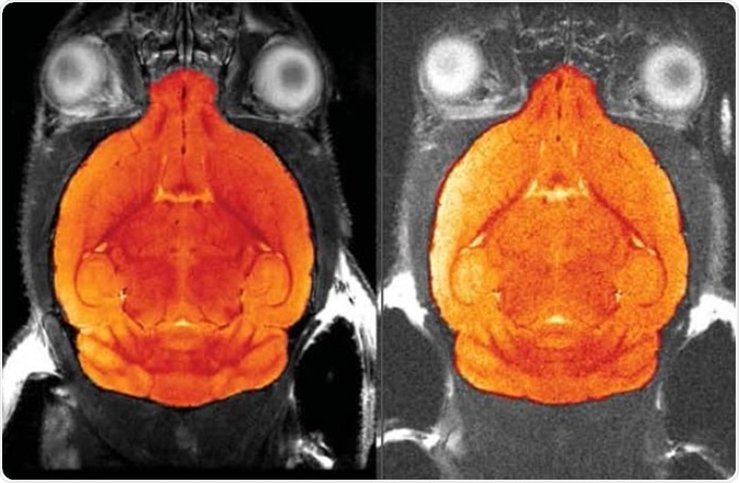
Comparison of 1H four channel phased array receive-only MRI CryoProbe with room temperature phased-array coil.
Mouse coronary artery imaging
Maximum intensity projection of a full mouse cardiac cycle allows for in vivo visualization of the coronary arteries of a mouse heart.
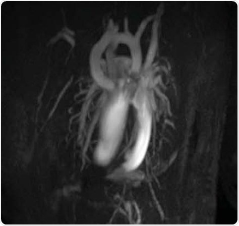
Mouse Coronary Artery lmaging.
1H MRS in mouse Hippocampus: 2.7µl voxel in vivo
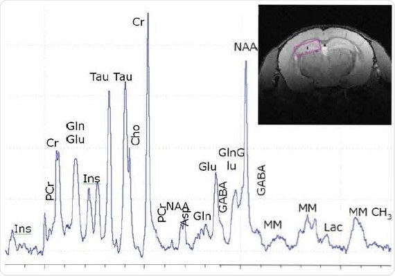
Localized 1H spectroscopy in mouse brain at 11.7 T in a 2.7µl voxel
Courtesy: M. Santin, R. Paquin et al., ICrv1-CENIR, Paris, France.
Mouse spine imaging
The below picture shows high-resolution (46 x 46 µm in-plane) mouse spine imaging acquired using TurboRARE, captured in less than 7 minutes at 9.4 T, clearly differentiating white and gray matter and revealing fine anatomical details, such as cerebrospinal fluid, vessels, and root ganglions.
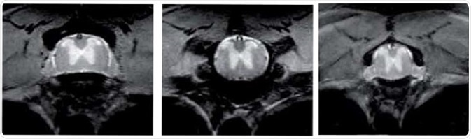
MouseSpine lmaging
The MRI CryoProbe family
1H four channel phased array receive-only MRI CryoProbe for 12 cm gradients and larger
The enormous signal gain helps to achieve a drastic reduction in scan time while maintaining a very high resolution. Using this MRI CryoProbe, a signal-to-noise-gain of 2.6 can be achieved when compared to a room temperature, 8 four channel phased array coil.
Bruker also offers four channel phased array coils at 7 T, 9.4 T and 11.7 T and can develop the four channel phased array coils at 4.7 T based upon request.
1H quadrature transmit/receive MRI CryoProbe for 6 cm gradients
This MRI CryoProbe is specially designed for the 11 cm mouse scanners and can also be employed in all BioSpec with a 6 cm gradient insert. Bruker also supplies this MRI CryoProbe as a commercial product at 11.7 T and 15.2 T.
X-nuclei MRI CryoProbe with combined 1H room temperature RF-coil
Lower NMR frequencies offer a number of signal-to-noise gain benefits. The SNR gain will be more for the nuclei if their frequency is low. Hence, signal-to-noise gains of a factor of 5 can be achieved in 13C measurements.
Bruker supplies this MRI CryoProbe as a commercial product at 7 T and 9.4 T and can develop other frequencies upon request.
DTI fibers
Brain connectivity studies are difficult to perform in small animals but can be achieved using sophisticated MRI technologies, such as a 7T BioSpec and MRI CryoProbe technology for image acquisition. The below picture shows high-resolution DT-MRI and fiber tracking of the live mouse brain, detailing the features of the fine cytoarchitecture of the nervous tissue and delineating the fiber tracts organization.
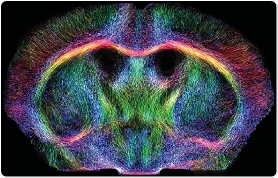
DTI fibers of the live mouse brain .Courtesy: L.-A. Harsan, D. von Elverfeldt et al., University Medical Center Freiburg, Freiburg, Germany.
Unmatched Results
ln vivo mouse brain with 19.5 µm resolution at 15.2 Tesla
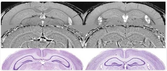
Comparison of micro-structures in the mouse brain measured at 15.2 T by using high resolution SWI with histological Nissle staining (below}.
Angiography
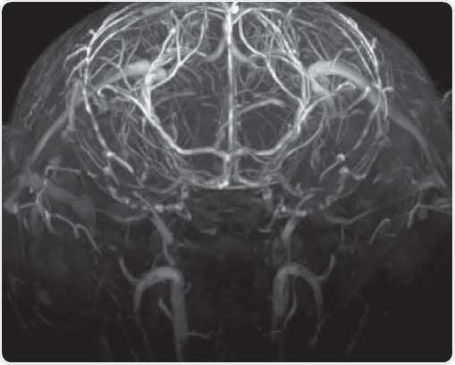
Time-of-flight angiography with no contrast agent at high spatial resolution showing excellent contrast enabling the identification of fine vascular structures
Testimonials
"This increased SNR can be invested in either reducing the acquisition time by a factor 6.25 at constant SNR or, as in the examples shown, in enhancing image quality, i.e. increase the CNR to improve the definition of anatomical structures. and/or to increase the spatial resolution in structural imaging ad angiography.”
Prof. Dr. Markus Rudin, ETH. Zürich. Switzerland
"ln conclusion, cardiac morphology, cardiac chamber quantification and cardiac function assessment using a cryogenically-cooled RF probe is feasible and affords SNR gains in the range of 3.0 to 5.0 compared to a conventional room temperature cardiac RF coil set-up.”
Prof. Dr. Thoralf Niendorf, MDC, Berlin, Germany
About Bruker
Bruker delivers the world's most comprehensive range of research tools enabling life science, materials science, analytical chemistry, process control and clinical research. Bruker is also the leading superconductor magnet and ultra high field magnet manufacturer for NMR and MRI solutions.
Sponsored Content Policy: News-Medical.net publishes articles and related content that may be derived from sources where we have existing commercial relationships, provided such content adds value to the core editorial ethos of News-Medical.Net which is to educate and inform site visitors interested in medical research, science, medical devices and treatments.
Last updated: Jul 28, 2017 at 7:18 AM


.png)


No hay comentarios:
Publicar un comentario