
New Method Pushes the Frontier in Imaging Resolution and Distinguishes Individual Features in Single Molecules
Published on August 3, 2017 at 10:19 AM
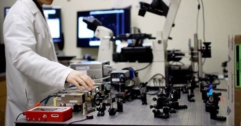
Image Credits: Andor Technology
Just as we are becoming familiar with super-resolution microscopes breaking the optical diffraction barrier to resolve details just 20 nm apart, scientists at Harvard University’s Wyss Institute for Biologically Inspired Engineering have demonstrated an optical resolution of less than five nanometres (<5 nm). The technology, which the team calls Discrete Molecular Imaging (DMI), was developed using either an ultra-sensitive Andor Zyla 4.2 or iXon Ultra 897 camera at its heart to achieve the highest resolution in optical microscopy yet seen.
Reporting in Nature Nanotechnology, Professor Peng Yin and his team detail DMI, an integrated set of new imaging methods that builds upon their DNA nanotechnology-powered super-resolution microscopy platform, DNA-PAINT. Because DMI can robustly image each individual molecular feature in a crowded molecular environment, it provides the capability for studying molecular conformations and heterogeneities in single multi-component complexes. It also complements current structural biology methods like X-ray crystallography and cryo-electron microscopy, and provides an easy, fast and multiplexed method for the structural analysis of many samples in parallel.
According to Professor Yin, “Proteins do not usually work in isolation but in larger complexes that enable cells to communicate with each other, move cargo around in their interiors, and replicate their DNA. Our ability to observe and track each individual protein within these machines is crucial to our ultimate understanding of these processes. The ultra-high resolution of DMI advances the DNA-PAINT platform one step further towards the vision of providing the ultimate view of biology.
The DNA-PAINT technologies developed by Professor Yin and his team are based on the transient binding of two complementary short DNA strands, one being attached to the molecular target that the researchers aim to visualize and the other attached to a fluorescent dye. Repeated cycles of binding and unbinding create a precisely defined blinking behaviour at the target site. The blinking is highly programmable by the choice of DNA strands and has now been further exploited by the team's current work to achieve ultra-high resolution imaging.


.png)


No hay comentarios:
Publicar un comentario