Viruses: Manufacturing Tycoons?

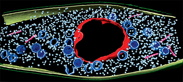
A computer image shows a bacterial cell invaded by a virus. The virus uses the cell to copy itself many times. It has built a protein compartment (red, rough circle surrounding the center) to house its DNA. Viral heads (blue, smaller pentagonal shapes spread through out) and tails (pink, rod shaped near the edges) are essential parts of a finished viral particle. The small, light blue particles are the bacterium’s own protein-making ribosomes. Credit: Vorrapon Chaikeeratisak, Kanika Khanna, Axel Brilot and Katrina Nguyen.
As inventors and factory owners learned during the Industrial Revolution, the best way to manufacture a lot of products is with an assembly line that follows a set of precisely organized steps employing many copies of identical and interchangeable parts. Some viruses are among life’s original mass producers: They use sophisticated organization principles to turn bacterial cells into virus particle factories.
Scientists at the University of California, San Diego, and the University of California, San Francisco, used cutting-edge techniques to watch a bacteria-infecting virus (bacteriophage) set up its particle-making factory inside a host cell.
The image above shows this happening inside a Pseudomonas chlororaphis, a soil-borne bacterium that protects plants against fungal pathogens. The virus builds a compartment (red, rough circle surrounding the center) that helps organize an assembly line for making copies of itself. The compartment looks like a cell nucleus, which bacteria do not have, and it functions like a nucleus by keeping activities that directly involve DNA separate from other cellular functions.
To understand the steps the virus takes to hijack and alter bacteria so effectively, the researchers stained viral proteins. This approach allowed them to watch the process through a light microscope. The nucleus-like compartment appears right after a virus particle infects the cell, and viral DNA gets copied inside it. Later, the proteins that make up the virus’ head begin to appear outside the compartment. When assembled, the viral heads dock beside the compartment and are filled with copies of viral DNA. Proteins that form a viral tail also appear outside the compartment and link up with the heads to produce a finished viral particle. The virus continues copying itself until there are enough virus particles to burst the cell open and kill it. The scientists confirmed their work using cryo-electron microscopy, an imaging technique that allowed them to look at bacteria in a near-natural state without the need for protein staining.
According to Joe Pogliano  , part of the research team, the level of sophistication the bacteriophage uses to build structures inside a cell to improve its copying ability is remarkable. The virus makes the bacterium function like a plant or animal (eukaryotic) cell, an action that has not been observed before. It may provide support to the theory of viral eukaryogenesis, which proposes that nucleus-containing eukaryotic cells like the ones that make up animals and plants could have evolved when a virus infected a bacterium over 2 billion years ago.
The researchers suggested that the virus may build the compartment to protect its vulnerable DNA from the bacterium’s defense systems. In doing so, the virus also creates a cellular factory with characteristics strikingly like those of more complex eukaryotic cells.
This work was funded in part by the National Institute of General Medical Sciences under grants R01GM031627, R01GM104556, R01GM057045 and DP2GM123494.


.png)


No hay comentarios:
Publicar un comentario