|
|
| | November 21, 2018 | |
| | |
| | The latest life science microscopy news from AZoNetwork | |
|
|
|
|
|
|
 |
| |
 Scientists have used cryo-electron microscopes to generate 3D images of amyloid, showing that aggregates form long, twisted fibers. Scientists have used cryo-electron microscopes to generate 3D images of amyloid, showing that aggregates form long, twisted fibers. | |
|
| |
 Fluorescent microscopy has revolutionized the study of bacteria and helped scientists to understand various aspects of their growth, development, and pathogenesis. Fluorescent microscopy has revolutionized the study of bacteria and helped scientists to understand various aspects of their growth, development, and pathogenesis. | |
|
| |
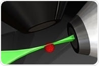
_a22f34786d3240f680c6b43ca3221bac-310x240__636783848949204550_multisizecolor_150_gray.jpg) Total internal reflection fluorescence (or TIRF) microscopy excites fluorophores in a thin region of the specimen. This way, only fluorescent molecules that are close to the solid (usually a glass coverslip) are efficiently excited. Total internal reflection fluorescence (or TIRF) microscopy excites fluorophores in a thin region of the specimen. This way, only fluorescent molecules that are close to the solid (usually a glass coverslip) are efficiently excited. | |
|
| |
 Correlative Light and Electron Microscopy, or ‘CLEM’, is an imaging approach that couples light microscopy’s varied capabilities with electron microscopy’s high resolution. One of the main disadvantages for biological uses of CLEM is the requirement to dry or cryogenically freeze samples, which makes experimental methods overly complex and has a tendency to introduce artifacts. Correlative Light and Electron Microscopy, or ‘CLEM’, is an imaging approach that couples light microscopy’s varied capabilities with electron microscopy’s high resolution. One of the main disadvantages for biological uses of CLEM is the requirement to dry or cryogenically freeze samples, which makes experimental methods overly complex and has a tendency to introduce artifacts. | |
|
|
|
|
|


.png)


No hay comentarios:
Publicar un comentario