What are Osteoclasts?
Osteoclasts are specialized, multinucleated cells which play a vital role in bone development and regeneration.
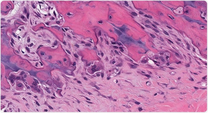 Credit: vetpathologist/Shutterstock.com
Bone is an important composite connective tissue which is remodeled continuously to repair damage, balance calcium levels in the body and maintain the integrity of the skeleton.
The synchronized response of the bone-forming cells (osteoblasts) and osteoclasts are responsible for the dynamic process of bone remodeling. Osteoclasts clear away the aged or damaged mineralized and calcified constituents of the bone matrix.
Formation and differentiation
Osteoclasts originate from the hematopoietic stem cells (HSC) under the presence of macrophage colony stimulating factor (MCSF) and receptor activator of NF-κB ligand (RANKL). The HSC produces a common myeloid progenitor cell which further differentiates into granulocyte/macrophage progenitor (GMP) under the influence of granulocyte/macrophage colony stimulating factor (GM-CSF).
MCSF stimulates the differentiating GMP into cells of macrophage or monocyte lineage which are established osteoclast precursors. The pre-osteoclast cells differentiate to form mature osteoclasts in the presence of RANKL, a member of the tumor necrosis family (TNF).
Osteoclasts are found in various subtype and differ based on the matrix they are positioned, the time of day, and the type of remodeling they perform.
Structural features
Osteoclasts consist of two distinct features; a ruffled border composed of convoluted membrane and a sealing zone which contains actin filament ring. The ruffled border develops by merging of secretory lysosomes with the plasma membrane. The actin ring surrounds the ruffled border and isolates the acidified microenvironment from the general extracellular space.
Function in bone resorption
Osteoclasts break down both the organic and inorganic components of the bone. They do this by secreting acid and various proteolytic enzymes.
The bone resorption process starts with the polarization of osteoclast leading to the formation of the bone-apposed ruffled membrane. The sealing zone forms an isolated microenvironment, enclosed by the cell and the surface of the bone. The ruffled membrane secretes protons through a vacuolar-type ATPase which combine with chloride transported through chloride channel.
The hydrochloric acid secreted in the resorption compartment dissolves the inorganic components of the underlying bone. After demineralization, the organic component of bone is eaten up by the lysosomal secreted proteolytic enzyme, cathepsin K along with matrix metalloprotease (MMP).
The degraded organic and inorganic materials are removed from the resorption compartment through endocytosis by the ruffled membrane. Osteoclasts may detach and reinitiate the resorption cycle or undergo death after completion of resorption. To maintain the normal physiological balance of bone function, they couple with osteoblasts.
After the degradation phase, osteoclasts promote formation and differentiation of the bone-forming cells. The osteoblasts outline the eroded bone and secrete the organic component of bone which is further mineralized. Certain growth factors such as insulin-like growth factor-1 (IGF-1) and transforming growth factor- β (TGF-β) influence the coupling between osteoblasts and osteoclast.
Pathological conditions
During normal bone remodeling, bone resorption is firmly coupled to the bone formation to ensure maintenance of bone mass and quality. Loss of the balanced coupling process or dysregulation in osteoclasts function and differentiation results in several bone diseases.
The following conditions demonstrate the importance of normal osteoclast function in the body:
- Osteoporosis is the most common bone disease, characterized by increased osteoclast activity. This increased activity leads to decrease in bone density resulting in fractures. Hormonal factors further influence the bone resorption process. The reduced level of estrogen in post-menopausal women increases osteoclast activity and reduces the deposition.
- Paget’s disease is a deforming bone disorder characterized by an increase in osteoclast number and size. The increase in the number of osteoclasts leads to localized bone destruction and compensatory activation of surrounding osteoblasts. The disease is caused by mutations in genes responsible for regulating the production of osteoclasts.
- Rheumatoid arthritis: bone destruction mediated by osteoclast are also observed in late stages of rheumatoid arthritis. The activation of pathological osteoclasts in the disease results in painful and erosive lesions.
- Osteopetrosis a rare bone disorder results from genetic mutations of the factor regulating osteoclast production or resorptive function leading to the accumulation of dense, brittle bones.
Reviewed by Kate Anderton, BSc
Sources:
- https://www.ncbi.nlm.nih.gov/pmc/articles/PMC4920143/
- https://www.ncbi.nlm.nih.gov/pubmed/20851921
- http://eknygos.lsmuni.lt/springer/61/01-23.pdf
- https://www.ncbi.nlm.nih.gov/pubmed/25008556
- https://www.ncbi.nlm.nih.gov/pmc/articles/PMC4882648/
- https://www.nature.com/articles/boneres20133.pdf?origin=ppub
- https://www.ncbi.nlm.nih.gov/pmc/articles/PMC1851862/
Further Reading
Last Updated: Mar 15, 2018


No hay comentarios:
Publicar un comentario