Snapshots of Life: Finding Where HIV Hides
Researchers have learned a tremendous amount about how the human immunodeficiency virus (HIV), which causes AIDS, infects immune cells. Much of that information comes from studying immune cells in the bloodstream of HIV-positive people. Less detailed is the picture of how HIV interacts with immune cells inside the lymph nodes, where the virus can hide.
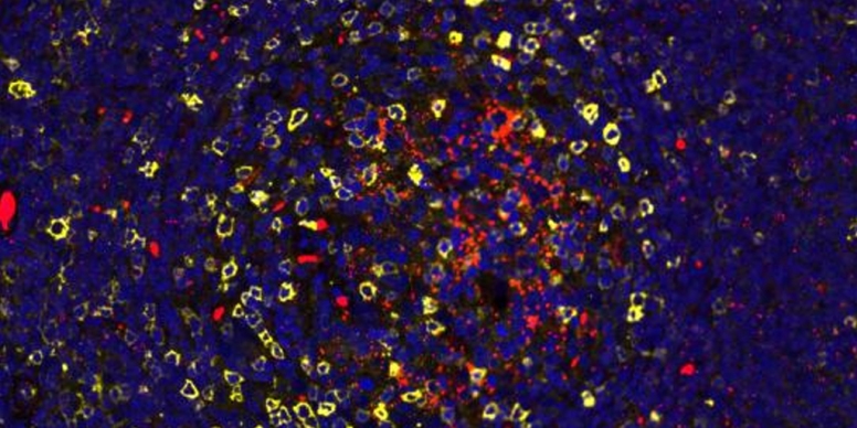
In this image of lymph tissue taken from the neck of a person with uncontrolled HIV infection, you can see areas where HIV is replicating (red) amid a sea of immune cells (blue dots). Areas of greatest HIV replication are associated with a high density of a subtype of human CD4 T-cells (yellow circles) that have been found to be especially susceptible to HIV infection.
This interesting micrograph was among the winners in the University of California, Berkeley (UC Berkeley) 2017 MIC Image Contest. It comes from the NIH-supported lab of Nadia Roan at the University of California, San Francisco (UCSF) and the J. David Gladstone Institutes, and was generated on a digitizing microscope slide scanner by Rajaa Hussien and imaged by Jen-Yi Lee at UC Berkeley.
The work is part of a larger effort in the Roan lab to understand which subtypes of CD4 T-cells are most vulnerable to HIV infection. Roan’s team initially performed these studies by infecting human cells with HIV in lab dishes. That work has led to some intriguing discoveries. As published recently, Roan and her colleagues identified another subtype of CD4 T-cells, characterized by another cell-surface protein called CD127. These cells allow HIV to enter, but with an unusual twist: once inside the cell, the virus can’t replicate for reasons that remain to be elucidated [1].
As Roan and others continue to learn more about HIV infection in lymph nodes and other types of tissue, they hope to discover important clues that may lead to new ways to prevent or control the infection. For instance, it might be possible to design treatments to target HIV in parts of the body where the virus likes to hide, and beautiful images like this one are helping researchers to find those hiding places.
Reference:
[1] Mass Cytometric Analysis of HIV Entry, Replication, and Remodeling in Tissue CD4+ T Cells. Cavrois M, Banerjee T, Mukherjee G, Raman N, Hussien R, Rodriguez BA, Vasquez J, Spitzer MH, Lazarus NH, Jones JJ, Ochsenbauer C, McCune JM, Butcher EC, Arvin AM, Sen N, Greene WC, Roan NR. Cell Rep. 2017 Jul 25;20(4):984-998.
Links:
HIV/AIDS (National Institute of Allergy and Infectious Diseases/NIH)
Nadia Roan (University of California, San Francisco; The J. David Gladstone Institutes)
NIH Support: National Institute of Allergy and Infectious Diseases


.png)


No hay comentarios:
Publicar un comentario