Volume 24, Number 3—March 2018
Dispatch
Whole-Genome Analysis of Mycobacterium tuberculosis from Patients with Tuberculous Spondylitis, Russia
On This Page
Ekaterina Chernyaeva , Mikhail Rotkevich, Ksenia Krasheninnikova, Andrey Yurchenko, Anna Vyazovaya, Igor Mokrousov, Natalia Solovieva, Viacheslav Zhuravlev, Piotr Yablonsky, and Stephen J. O’Brien
Abstract
Whole-genome analysis of Mycobacterium tuberculosis isolates collected in Russia (N = 71) from patients with tuberculous spondylitis supports a detailed characterization of pathogen strain distributions and drug resistance phenotype, plus distinguished occurrence and association of known resistance mutations. We identify known and novel genome determinants related to bacterial virulence, pathogenicity, and drug resistance.
Tuberculosis (TB) is an infectious disease caused by Mycobacterium tuberculosis, which typically affects the lungs but can affect other sites. In 2016, an estimated 10.4 million new TB cases and 1.6 million TB-related deaths were documented worldwide (1). The Russian Federation reported >120,000 TB cases and ≈13,700 TB deaths in 2016 (1). TB strains with multidrug resistance (MDR TB), characterized by resistance to isoniazid or rifampin, are common in the Russian Federation. The estimated rate of MDR TB was 27% among TB case-patients newly diagnosed in 2016 and 65% among previously treated case-patients in 2016 (1). Most TB cases are associated with pulmonary localization of the disease; however, in some cases, extrapulmonary TB develops. In Russia, the rate of extrapulmonary TB cases among new TB cases was 3.3% in 2014; most extrapulmonary TB cases are osteoarticular and genitourinary (2). Approximately 70% of osteoarticular TB cases are tuberculous spondylitis (TBS), which cause severe specific lesions of >1 components of the spine (2). We report whole-genome sequencing (WGS) and variant analyses of M. tuberculosis isolates from patients treated in Russia for TBS during 2007–2014.

Figure 1. Distribution of Mycobacterium tuberculosis isolates randomly collected from 71 patients with tuberculous spondylitis who received treatment at clinics of Research Institute of Phthisiopulmonology in 32 regions of the the Russian Federation, 2007–2014
The isolates were randomly collected from 71 TBS patients who received treatment at clinics of the Research Institute of Phthisiopulmonology in 32 regions of the Russian Federation (Figure 1). In these cases, M. tuberculosis isolates were cultured from extrapulmonary clinical material and stored. We assessed the susceptibility of these stored isolates to streptomycin, isoniazid, rifampin, ethambutol, pyrazinamide, ethionamide, ofloxacin, kanamycin, amikacin, cycloserine, capreomycin, and paraaminosalicylic acid according to World Health Organization recommendations (3). We isolated genomic DNA from cultured bacteria by using phenol/chloroform extraction and subjected bacterial DNA to WGS by using the MiSeq platform (Illumina, San Diego, CA, USA) to a mean coverage of 47× (range 18×–170×), covering ≥99% of the M. tuberculosis H37Rv reference genome (GenBank accession no. NC_000962.3). We deposited WGS reads in the NCBI Sequence Read Archive (accession no. PRJNA352769).
We aligned sequenced reads to the reference genome and called variants (single-nucleotide polymorphisms [SNPs] and short insertions/deletions) by using bioinformatics software: bowtie2 (http://bowtie-bio.sourceforge.net/bowtie2/index.shtml); SAMtools (http://samtools.sourceforge.net); VCFtools (http://vcftools.sourceforge.net); and FreeBayes (https://github.com/ekg/freebayes). We used mutations that had q-scores ≥20 for comprehensive analysis. We used concatenated SNPs for phylogenetic analysis by using the GTRCAT (general time-reversible model with rate heterogeneity accommodated by using discrete rate categories) maximum-likelihood algorithm from the RAxML software package (4) to calculate an approximation model and 100 bootstrap replications. To avoid misalignments, we annotated SNPs in repetitive genome regions and in genes encoding proteins that contain proline-glutamate or proline-proline-glutamate motifs and filtered them from analysis. We used PhyTB (5) and SpoTyping tools (6) for phylogenetic classification of M. tuberculosis genomes and verified SpoTyping output by using previously conducted conventional spoligotyping analysis for 20 isolates that were previously described (7).

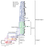
Figure 2. Phylogenetic analysis of Mycobacterium tuberculosis isolates obtained from patients with tuberculous spondylitis, Russian Federation, 2007–2014. Symbols indicate drug resistance: susceptible to all tested drugs; monoresistant to 1 drug; polyresistant, resistant to...
We identified 2 principal phylogenetic lineages among M. tuberculosis isolates, lineage 2 and lineage 4; further, we detected evolutionary ancient and modern sublineages within major lineage 2 (Beijing; Figure 2) according to previously described classifications (8). The largest subgroup within the Beijing clade belonged to the B0/W148 clonal cluster (Figure 2) (8). Lineage 4 was represented by 4 genetic families: Ural, 4.2; Latin-American/Mediterranean (LAM),(4.3); and T, 4.1 and 4.8. The 58 Beijing genotype isolates contained 38 MDR (65.5%) 5 extensively drug-resistant (XDR; resistant to isoniazid and rifampin plus any fluoroquinolone and >1 of 3 injectable second-line drugs) (8.6%), 7 polyresistant but not MDR (12%), 1 monoresistant (1.9%), and 7 susceptible (12%) TB isolates. The MDR TB frequency in the Beijing group (65.5%), was higher than that for other genetic groups pooled (p<0.0096 by Fisher exact test). The M. tuberculosis Beijing B0/W148 cluster was represented by 1 susceptible (4.8%), 2 polyresistant (9.5%), 3 XDR (14.3%), and 15 MDR (71.4%) TB isolates. The B0/W148 genetic group demonstrates an association with MDR TB (p = 0.03), shown previously (9,10). The other genetic groups (T, LAM, and Ural) included too few isolates to test for association with MDR TB.
Specimens of 50 TBS patients were HIV negative; 21 were HIV positive. Although we found no significant association of M. tuberculosis genetic groups to HIV infection, 42% of patients infected by B0/W148 strains were HIV positive whereas among patients infected by non-B0/W148 Beijing strains, only 22% were HIV positive. Further, only 14% patients infected with non-Beijing M. tuberculosis strains were HIV positive (Technical Appendix[PDF - 498 KB - 5 pages] Table 1).
We examined M. tuberculosis isolates for the presence of published variants associated with resistance to TB drugs (Table). We found a high level of concordance of phenotypic and genetic data for reported isoniazid- and rifampin-resistant isolates. We detected mutations in rpsL, gid, and rrs genes in 96.4% of streptomycin-resistant isolates; 81.8% of ofloxacin-resistant isolates had mutations in gyrA gene (there were no mutations in gyrB gene). Most ethambutol-resistant isolates (72.7%) showed mutations in the embA promoter region or embB region between codons 296 and 497. However, 3 ethambutol-susceptible isolates had mutations M306I (n = 1) and G406A (n = 2) in the embB gene. We detected mutations in genes pncA (11) and rpsA, associated with pyrazinamide resistance in 55.6% of pyrazinamide-resistant isolates, and 55% of kanamycin-resistant strains had mutations in the eis promoter. We detected no mutations in alr and ddl genes among cycloserine-resistant isolates, nor in thyA gene among paraaminosalicylic acid–resistant isolates.
Our analysis for small insertions and deletions detected 15 and 9, respectively, among the Beijing group (Technical Appendix[PDF - 498 KB - 5 pages] Table 2). A deletion in kdpD and an insertion in Rv1258c were previously described (12,13). The other 22 mutations are novel: 18 were specific to the Beijing group; 2 to the modern Beijing group; 1 to the ancient Beijing group; and 3 to the B0/W148 group. We identified most mutations in genes encoding membrane-associated proteins, although several mutations were in regulatory genes, genes involved in cell metabolism, probable transposase genes, and genes with unknown function.
One insertion and 2 deletions were significantly associated with B0/W148 genetic group in kdpD, mmr and Rv1995 (p = 2.5 × 10⁻¹⁷) genes. Merker et al. (12), who proposed a pathogenic influence for B0/W148 strains, described a frameshift deletion in kdpD among Beijing B0/W148 strains. Deletion in the kdpD gene can lead to the formation of nonfunctional proteins KdpD and KdpE. Parish et al. showed that M. tuberculosis lacking KdpD and KdpE function express increased virulence in a mouse model of infection (14), which supports that the kdpD deletion detected in our study may influence Beijing B0/W148 strain’s rapid expansion and virulence. A mutation in the promoter region of the mmr gene (Rv3065), encoding multidrug-transport integral membrane protein, might contribute to drug resistance in Beijing B0/W148 strains. Sriraman et al. recently showed that mmr is upregulated in rifampin-resistant and MDR TB strains, even in the presence of target gene mutations (15). Insertion in Rv1258c is common to all Beijing strains except ancient TB0010. In their study, Villellas et al. found the cytosine nucleotide insertion between positions 580 and 581 in the Rv1258c gene in all Beijing isolates among streptomycin-resistant M. tuberculosis strains (13).
In conclusion, we examined the phylogenetic and drug-resistance properties of M. tuberculosis isolates collected from 71 TBS patients in 32 locales across Russia. Our analyses confirmed the phylogenetic separation of pathogenic M. tuberculosis strains and support the prevalence of Beijing strains showing high levels of multidrug resistance among TBS isolates. Further, we found known SNP variants that had high concordance with suggested drug resistance. Finally, novel insertions/deletions were apparent, which we suggest are candidates for conferring drug resistance pending independent replication studies. Our analysis of WGS data identified known and novel genetic determinants that could or do influence bacterial virulence, pathogenicity, and drug resistance.
Dr. Chernyaeva is a senior researcher at Theodosius Dobzhansky Center for Genome Bioinformatics at St. Petersburg State University, St. Petersburg, Russia. Her main research focuses on M. tuberculosis comparative genomics to discover genetic markers associated with microbiological and clinical features.
References
Acknowledgments
This work was supported by the Russian Foundation for Basic Research (grant 16-34-60163) and St. Petersburg State University (grants 1.38.253.2015 and 1.52.1647.2016).
E.C., V.Z., and S.O.B. designed the experiment. N.S., V.Z., and P.Y. provided M. tuberculosis genomic DNA, information on drug resistance, and clinical data. E.C. performed whole-genome sequencing. E.C., M.R., K.K., and A.Y. performed bioinformatics and statistical analysis of whole-genome sequencing data. A.V. and I.M. conducted M. tuberculosis spoligotyping. E.C., I.M., and S.O.B. wrote the manuscript.
References
- World Health Organization. Global tuberculosis report 2017. Geneva: The Organization; 2017 [cited: Nov. 27, 2017]. http://apps.who.int/iris/bitstream/10665/259366/1/9789241565516-eng.pdf?ua=1
- Yablonsky P, Mushkin A, Belilovsky E, Galkin V. Extrapulmonary tuberculosis. In: Tuberculosis in the Russian Federation 2012/2013/2014 [in Russian]. Moscow. Triada; 2015; 129–35 [cited Nov. 27, 2017].
- World Health Organization. Guidelines for surveillance of drug resistance in tuberculosis. 4th Edition. Geneva: The Organization; 2009.
- Stamatakis A. RAxML-VI-HPC: maximum likelihood-based phylogenetic analyses with thousands of taxa and mixed models. Bioinformatics. 2006;22:2688–90. DOIPubMed
- Benavente ED, Coll F, Furnham N, McNerney R, Glynn JR, Campino S, et al. PhyTB: Phylogenetic tree visualisation and sample positioning for M. tuberculosis. BMC Bioinformatics. 2015;16:155. DOIPubMed
- Xia E, Teo YY, Ong RT. SpoTyping: fast and accurate in silico Mycobacterium spoligotyping from sequence reads. Genome Med. 2016;8:19. DOIPubMed
- Kamerbeek J, Schouls L, Kolk A, van Agterveld M, van Soolingen D, Kuijper S, et al. Simultaneous detection and strain differentiation of Mycobacterium tuberculosis for diagnosis and epidemiology. J Clin Microbiol. 1997;35:907–14.PubMed
- Mokrousov I. Insights into the origin, emergence, and current spread of a successful Russian clone of Mycobacterium tuberculosis. Clin Microbiol Rev. 2013;26:342–60. DOIPubMed
- Vyazovaya A, Mokrousov I, Solovieva N, Mushkin A, Manicheva O, Vishnevsky B, et al. Tuberculous spondylitis in Russia and prominent role of multidrug-resistant clone Mycobacterium tuberculosis Beijing B0/W148. Antimicrob Agents Chemother. 2015;59:2349–57. DOIPubMed
- Lasunskaia E, Ribeiro SC, Manicheva O, Gomes LL, Suffys PN, Mokrousov I, et al. Emerging multidrug resistant Mycobacterium tuberculosis strains of the Beijing genotype circulating in Russia express a pattern of biological properties associated with enhanced virulence. Microbes Infect. 2010;12:467–75. DOIPubMed
- Yadon AN, Maharaj K, Adamson JH, Lai YP, Sacchettini JC, Ioerger TR, et al. A comprehensive characterization of PncA polymorphisms that confer resistance to pyrazinamide. Nat Commun. 2017;8:588. DOIPubMed
- Merker M, Blin C, Mona S, Duforet-Frebourg N, Lecher S, Willery E, et al. Evolutionary history and global spread of the Mycobacterium tuberculosis Beijing lineage. Nat Genet. 2015;47:242–9. DOIPubMed
- Villellas C, Aristimuño L, Vitoria MA, Prat C, Blanco S, García de Viedma D, et al. Analysis of mutations in streptomycin-resistant strains reveals a simple and reliable genetic marker for identification of the Mycobacterium tuberculosis Beijing genotype. J Clin Microbiol. 2013;51:2124–30. DOIPubMed
- Parish T, Smith DA, Kendall S, Casali N, Bancroft GJ, Stoker NG. Deletion of two-component regulatory systems increases the virulence of Mycobacterium tuberculosis. Infect Immun. 2003;71:1134–40. DOIPubMed
- Sriraman K, Nilgiriwala K, Saranath D, Chatterjee A, Mistry N. Deregulation of genes associated with alternate drug resistance mechanisms in Mycobacterium tuberculosis. Curr Microbiol. 2017. DOIPubMed


.png)


No hay comentarios:
Publicar un comentario