
You’ve Got Questions, We’ve Got Answers: Cell Day 2016

Students from Connecticut to Washington State and points in between peppered our experts with questions during the recent live Cell Day web chat. They fielded questions about cell structures, microscopes and other tools, life as a scientist, and whether there are still discoveries to be made in cell biology. One of the Cell Day moderators, Jessica Faupel-Badger, even gave a shout-out to the Biomedical Beat blog as a great way to keep up with new and exciting discoveries being made every day. Thanks!
The full transcript with all the questions and answers is now available. We’ve recapped some of the highlights below.
[Check out our Facebook Live post-chat video  for a bonus answer to the question “If you put lizard DNA into human cells, could humans regrow their limbs?”]
Being a Scientist

Prior to joining NIGMS in 2016 as a program director, Patrick Brown, was a high school chemistry teacher in Maryland.
What do you think is the best thing about being a biologist? Why do you love your job so much? (Assuming you do!)
Patrick Brown answered: I love that question! And, I love being a scientist. There are so many things that I like about my career choice. The answer is simple—I like learning! I like learning about different living organisms and how they may be the same or different. I also really enjoy the multi-cultural aspect of science. I get to interact with so many different people from different parts of the world who are all studying different aspects of science that are just as interesting as my own, and we are all interested in knowing more about life.
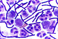
Gram stained cerebrospinal fluid with gram-positive anthrax bacilli. Credit: Wikimedia Commons, Yuval Madar.
How did you know that biology was the career for you? In other words, what motivated you to become a biologist?
Amy Kullas answered: I remember being in my high school biology class, gazing through a microscope, and seeing the mixture of beautiful purple and pink cocci after performing my first Gram stain . It was at that moment that I got hooked on science. I majored in microbiology in college and then went on to graduate school.
What does a typical day at work look like?
 Amicia Elliott answered: The truth is that every day at work is an adventure. A typical day includes some of the following things: reading scientific papers, thinking about and designing experiments (my favorite part!), carrying out those experiments, data analysis and discussing results. Scientists work long hours to accomplish all of these things, but it is mostly a labor of love!
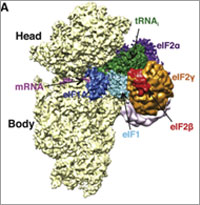
In a 2014 Molecular Cell paper, NIGMS Director Jon Lorsch and colleagues determined the structure of initiation complexes.
What was the most interesting experiment you have conducted?
Jon Lorsch answered: In my lab, we study how proteins are synthesized by the eukaryotic ribosome. We have learned a great deal about how the ribosome and the proteins that help it (called translation factors) find the start codon in the messenger RNA. Recently, in collaboration with a group in the UK, we used cryo-electron microscopy to determine the three-dimensional structure of various ‘initiation complexes’ – the small subunit of the ribosome bound to mRNA, tRNA and initiation factors. Being able to see how this process works in three dimensions is amazing!
Inside the Cell
What is a cell?
 Joe Gindhart answered: That is a deep question! In a biological context, a cell is the basic unit of life. As Rudolf Virchow wrote, ‘Omnis cellula e cellula,’ which means all cells come from pre-existing cells. Cells were first observed in the 1600s by Robert Hooke, who used an early microscope to look at bark from a cork tree
What are the biggest and smallest cells?
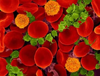
Human red blood cells. Credit: Dennis Kunkel Microscopy, Inc.
Dorit Zuk answered: That’s a good question because it depends on how you measure. For example, sperm cells have a diameter of 1 to 3 micrometers, but a tail of 50 micrometers. On the other hand, the human red blood cell has a diameter of 4 to 8 micrometers so may be considered the smallest. In humans, the largest cell in diameter (across) is the egg cell, which is about the size of the period at the end of this sentence. Some of the longest cells in the body are nerve cells—they can be over 3 feet long.
How long do cells live and what happens to them when they die?
Tracy Koretsky answered: Many cells in the body are constantly turning over. Cells of the small intestine only survive 2 to 4 days. Skin cells turn over every 10 to 30 days. Nerve cells stop dividing when fully differentiated. So, some cells are constantly dying and new cells are created by cell division whereas other cells are not replaced after they die. In multicellular organisms with immune systems, dead cells are cleared from the body by white blood cells called macrophages, which engulf and destroy the dead cells inside cell organelles called lysosomes. This process is called phagocytosis.
Which organelle does the most moving inside of a cell? Is there a way to measure how fast this organelle moves?
Shireen Sarraf answered: The inside of the cell is a very dynamic environment. Proteins and biochemical signal molecules need to move all over the cell to do their jobs. These proteins and molecules are often delivered by vesicles, which are membrane-bound organelles that often originate at the Golgi apparatus and then travel through the cell. In some cases, the cellular products are released outside the cell across the plasma membrane. This phenomenon has a key role in synaptic neurotransmission, endocrine secretion, mucous secretion, and other processes. Scientists can measure vesicle movement by using microscopes to track vesicles labeled with fluorescent dyes.
New Technologies Bring New Discoveries
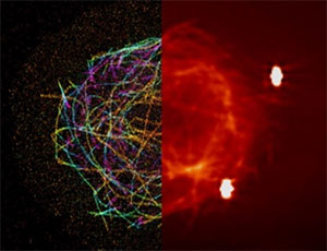
A super-resolution image (left) of the molecular scaffolding, called microtubules, in the cell of a fruit fly, combined with a conventional microscopy image (right) of the same cell for comparison. Credit: Jim and Cathy Galbraith, Glob Shtengel, Harald Hoss/HHMI/Janelia Research Campus.
What is the best microscope to view cells?
Alex Valm answered: There are many different kinds of microscopes that have specific qualities that make them best for answering different kinds of questions. Generally, there are two main kinds of microscopes: electron microscopes and light microscopes. Electron microscopes allow the greatest magnification and resolution of small objects–down to the scale of molecules, but they have two major limitations: First, they can only be used on dead cells and, second, they cannot be used with labels to identify specific parts of the cell. Light microscopes have lower resolution, but they can be used on living cells. Fluorescence microscopy allows scientists to label specific proteins or other structures in the cell and recent advances in light microscopy allow super resolution, the ability to see objects smaller than the light microscope resolution limit. The 2014 Nobel Prize in Chemistry was given to the inventors of super-resolution microscopy.
How can cells be so powerful even though they are very small?
 Alison Cole answered: That’s a very interesting question and there are lots of ways to answer it. One way is to say that even though they are tiny, cells can grow and reproduce because they contain DNA—the hereditary material of genes—and RNA, which contains the information necessary to build various proteins such as enzymes, the cell’s primary machinery. All cells (except red blood cells which lack a cell nucleus) possess this hereditary material. It’s amazing that so much genetic material can fit into these tiny cells!
Jessica Faupel-Badger was one of the Cell Day moderators.
What are some new exciting discoveries that have been made about cells recently?
Jessica Faupel-Badger answered: Cell biology is a very broad field, and each person in the room might have a different answer to this question based on their own interests. One function of the cell that you may not be aware of is autophagy, the process of the cell recycling and degrading intracellular components. Autophagy was the subject of the 2016 Nobel Prize in Physiology or Medicine .
More to Explore
There’s so much to discover in the vast, tiny world of the cell. Find more about cell biology, genetics, chemistry and structural biology as well as science careers in NIGMS’ science education materials.
Here a few good reads to get you started:
- Go deep inside the cell with this comprehensive tour.
- Is being a scientist the best job ever? These people think so!
- Scientists have many ways to see the invisible.
- Can’t get enough about cell organelles? Textbooks may need to be updated, thanks to these new findings.


.png)

.png)


No hay comentarios:
Publicar un comentario