
Volume 23, Number 1—January 2017
Letter
Puumala Virus in Bank Voles, Lithuania
On This Page
Abstract
Little is known about the presence of human pathogenic Puumala virus (PUUV) in Lithuania. We detected this virus in bank voles (Myodes glareolus) in a region of this country in which previously PUUV-seropositive humans were identified. Our results are consistent with heterogeneous distributions of PUUV in other countries in Europe.
Puumala virus (PUUV) (family Bunyaviridae) is an enveloped hantavirus that contains a single-stranded trisegmented RNA genome of negative polarity (1). PUUV harbored by the bank vole (Myodes glareolus) is the most prevalent human pathogenic hantavirus in Europe (2). A high population density of bank voles can lead to disease clusters and possible outbreaks of nephropathia epidemica, a mild-to-moderate form of hantavirus disease (3).
In contrast to the Fennoscandian Peninsula and parts of central Europe (4,5), little is known about the epidemiology of PUUV in Poland and the Baltic States. Recent investigations confirmed the presence of PUUV in certain parts of Poland (5,6). A molecular study of bank voles in Latvia identified 2 PUUV lineages (Russian and Latvian) (7). In Estonia, serologic and molecular screening provided evidence of the Russian PUUV lineage (8). For Lithuania, a previous serosurvey indicated the presence of PUUV-specific antibodies in humans from 3 counties (Technical Appendix[PDF - 329 KB - 4 pages]Figure 1). However, molecular evidence of PUUV in humans or in voles is lacking (9).
We report a molecular survey of rodent populations in Lithuania at 5 trapping sites, including 2 sites in counties where PUUV-specific antibodies were previously detected in humans (Technical Appendix[PDF - 329 KB - 4 pages]Figure 1). A total of 134 bank voles, 72 striped field mice (Apodemus agrarius), and 59 yellow-necked field mice (A. flavicollis) were captured during 2015. Three trapping sites (Juodkrantė, Elektrėnai, and Lukštas) were located in forests at or near a cormorant colony, and 2 trapping sites (Žalgiriai and Rusnė) were located in a wet forest and flooded meadows. All applicable institutional and national guidelines for the care and use of animals were followed.
For PUUV detection, we extracted RNA from bank vole lung tissue samples by using the Qiazol Protocol (QIAGEN, Hilden, Germany) and conducting screening by using a small segment RNA–specific reverse transcription PCR (RT-PCR) and primers Pu342F and Pu1102R (6). We detected PCR products for 5 (LT15/164, LT15/165, LT15/166, LT15/174, and LT15/201) of 45 bank voles from the Lukštas trapping site. All 9 striped field mice and 2 yellow-necked field mice from Lukštas showed negative results for the PUUV RT-PCR.
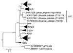
Figure. Phylogenetic tree based on complete nucleocapsid gene sequences of Puumala virus (PUUV) strains from Lithuania (LT), Latvia (Jelgava1), and other PUUV clades. Tula virus (TULV) was used as the outgroup. The...
We amplified the complete nucleocapsid protein–encoding region for 3 of the 5 samples positive by RT-PCR with 3 primer pairs: PuNCRS (5′-TAGTAGTAGACTCCTTGAA-3′)/Pu255R (5′-TGGACACAGCATCTGCCA-3′), Pu40F (5′-CTGGAATGAGTGACTTAAC-3′)/Pu393R (5′-TATGGTAATGTCCTTGATGT-3′), and Pu1027F (5′-ATGGCAGAGTTAGGTGCA-3′)/Pu1779R (5′-TCAGCATGTTGAGGTAGT-3′). RT-PCR products were directly sequenced by using the BigDye Terminator Version 1.1 Cycle Sequencing Kit (Applied Biosystems, Darmstadt, Germany). We deposited the sequences of the 5 samples in GenBank under accession nos. KX757839, KY757840, KX 757841, KX751706, and KX751707 (Figure; Technical Appendix[PDF - 329 KB - 4 pages] Figure 2).
The 3 nucleocapsid protein–encoding nucleotide sequences showed identities of 98.2%–99.8%, and the 3 deduced nucleocapsid protein amino acid sequences showed identities of 99.8%–100% (Technical Appendix[PDF - 329 KB - 4 pages] Table). We found the highest similarity of the 3 nucleotide and corresponding amino acid sequences for the PUUV strain from Latvia (Jelgava1/Mg149/2008; JN657228): nucleotide sequence 89.8%–90.4% and amino acid sequence 99.8%–100% (Technical Appendix[PDF - 329 KB - 4 pages] Table).
We generated phylogenetic trees by using MrBayes 3.2.6 software (http://mrbayes.sourceforge.net/download.php) and MEGA6 software (http://www.megasoftware.net/) for complete (1,302 nt; Figure) and partial (465 nt; Technical Appendix[PDF - 329 KB - 4 pages] Figure 2) nucleocapsid protein–encoding sequences. Phylogenetic analysis confirmed results of pairwise nucleotide sequence divergence analysis, which indicated clustering of PUUV sequences from Lithuania with sequences from northern Poland (Technical Appendix[PDF - 329 KB - 4 pages] Figure 2) and the Jelgava 1 strain from Latvia (Figure). These sequences of the Latvian clade are well separated from the Russian and all other European PUUV clades.
To evaluate a potential association of PUUV with evolutionary lineages of the bank vole, we determined vole cytochrome b gene sequences, deposited them in GenBank under accession nos. KX769843 (LT15/164), KX769844 (LT15/165), KX769845 (LT15/166), KX769846 (LT15/174), and KX769847 (LT15/201), and compared them with cytochrome b prototype sequences of evolutionary lineages. Consistent with results for northern Poland (6), we identified 2 bank vole lineages at Lukštas, and the PUUV sequences were detected in 4 bank voles of the Carpathian phylogroup and in 1 vole of the Eastern lineage.
In conclusion, we detected PUUV in bank voles at 1 site (Lukštas) in Lithuania (prevalence of 11.1%). This site is located in a region where PUUV-seropositive persons were identified (9) and near the border with Latvia (Technical Appendix[PDF - 329 KB - 4 pages] Figure 1). The absence of PUUV in bank voles at 4 other sites might have been caused by the small number of voles tested. However, our results are consistent with heterogeneous distributions of PUUV in other countries (10).
Detection of this novel PUUV strain by using a specific RT-PCR confirms the reliability of this assay for molecular diagnostic and epidemiologic studies of this virus in Lithuania. Future large-scale monitoring studies are needed to evaluate the geographic distribution and temporal fluctuation of PUUV in bank vole populations in Lithuania.
Ms. Straková is a doctoral student at Masaryk University, Brno, Czech Republic. Her research interests are zoonotic viruses, vectorborne diseases, and molecular diagnostics.
Acknowledgments
We thank Nicole Reimer for generating Technical Appendix Figure 1.
P.S. was supported by a stipend from the Erasmus Programme.
References
- Plyusnin A, Beaty BJ, Elliot RM, Goldbach R, Kormelink R, Lundkvist A, et al. Family Bunyaviridae. In: King AM, Adams MJ, Carstens EB, Lefkowitz EJ, editors. Virus taxonomy: ninth report of the international committee on taxonomy of viruses. San Diego: Elsevier Academic Press; 2012. p. 725–41.
- Heyman P, Ceianu CS, Christova I, Tordo N, Beersma M, João Alves M, et al. A five-year perspective on the situation of haemorrhagic fever with renal syndrome and status of the hantavirus reservoirs in Europe, 2005-2010. Euro Surveill. 2011;16:19961.PubMed
- Clement J, Maes P, van Ypersele de Strihou C, van der Groen G, Barrios JM, Verstraeten WW, et al. Beechnuts and outbreaks of nephropathia epidemica (NE): of mast, mice and men. Nephrol Dial Transplant. 2010;25:1740–6. DOIPubMed
- Klempa B, Radosa L, Krüger DH. The broad spectrum of hantaviruses and their hosts in Central Europe. Acta Virol. 2013;57:130–7. DOIPubMed
- Michalski A, Niemcewicz M, Bielawska-Drózd A, Nowakowska A, Gaweł J, Pitucha G, et al. Surveillance of hantaviruses in Poland: a study of animal reservoirs and human hantavirus disease in Subcarpathia. Vector Borne Zoonotic Dis. 2014;14:514–22. DOIPubMed
- Ali HS, Drewes S, Sadowska ET, Mikowska M, Groschup MH, Heckel G, et al. First molecular evidence for Puumala hantavirus in Poland. Viruses. 2014;6:340–53. DOIPubMed
- Razzauti M, Plyusnina A, Niemimaa J, Henttonen H, Plyusnin A. Co-circulation of two Puumala hantavirus lineages in Latvia: a Russian lineage described previously and a novel Latvian lineage. J Med Virol. 2012;84:314–8. DOIPubMed
- Golovljova I, Sjölander KB, Lindegren G, Vene S, Vasilenko V, Plyusnin A, et al. Hantaviruses in Estonia. J Med Virol. 2002;68:589–98. DOIPubMed
- Sandmann S, Meisel H, Razanskiene A, Wolbert A, Pohl B, Krüger DH, et al. Detection of human hantavirus infections in Lithuania. Infection. 2005;33:66–72. DOIPubMed
- Drewes S, Turni H, Rosenfeld UM, Obiegala A, Strakova P, Imholt C, et al. Reservoir-driven heterogeneous distribution of recorded human Puumala virus cases in South-West Germany. Zoonoses Public Health. 2016. In press.


.png)


No hay comentarios:
Publicar un comentario