Snapshots of Life: Lighting up the Promise of Retinal Gene Therapy | NIH Director's Blog
Snapshots of Life: Lighting up the Promise of Retinal Gene Therapy
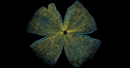
Caption: Large-scale mosaic confocal micrograph showing expression of a marker gene (yellow) transferred by gene therapy techniques into the ganglion cells (blue) of a mouse retina.
Credit: Keunyoung Kim, Wonkyu Ju, and Mark Ellisman, National Center for Microscopy and Imaging Research, University of California, San Diego
Credit: Keunyoung Kim, Wonkyu Ju, and Mark Ellisman, National Center for Microscopy and Imaging Research, University of California, San Diego
The retina, like this one from a mouse that is flattened out and captured in a beautiful image, is a thin tissue that lines the back of the eye. Although only about the size of a postage stamp, the retina contains more than 100 distinct cell types that are organized into multiple information-processing layers. These layers work together to absorb light and translate it into electrical signals that stream via the optic nerve to the brain.
In people with inherited disorders in which the retina degenerates, an altered gene somewhere within this nexus of cells progressively robs them of their sight. This has led to a number of human clinical trials—with some encouraging progress being reported for at least one condition, Leber congenital amaurosis—that are transferring a normal version of the affected gene into retinal cells in hopes of restoring lost vision.
To better understand and improve this potential therapeutic strategy, researchers are gauging the efficiency of gene transfer into the retina via an imaging technique called large-scale mosaic confocal microscopy, which computationally assembles many small, high-resolution images in a way similar to Google Earth. In the example you see above, NIH-supported researchers Wonkyu Ju, Mark Ellisman, and their colleagues at the University of California, San Diego, engineered adeno-associated virus serotype 2 (AAV2) to deliver a dummy gene tagged with a fluorescent marker (yellow) into the ganglion cells (blue) of a mouse retina. Two months after AAV-mediated gene delivery, yellow had overlaid most of the blue, indicating the dummy gene had been selectively transferred into retinal ganglion cells at a high rate of efficiency [1].
The researchers also used AAV2 to deliver into the retinas of mice a gene that coded for a mutant version of a protein, called DRP1. They found it inhibited normal DRP1 protein and stopped retinal ganglion cells from dying in response to glaucoma, a vision-threatening condition that elevates pressure within the eye. The gene transfer proved successful in rescuing the retinal ganglion cells, and researchers are continuing to pursue this line of study with the aim of translating their discoveries into possible ways of helping humans with glaucoma.
It’s also worth noting that this image recently took top honors in the NIH Institute and Centers Art Challenge, which was held in October as part of the Combined Federal Campaign, in which federal employees make their annual contributions to charitable organizations. The art competition had a number of outstanding entrants, some of which will be on display during November at the NIH Clinical Center in Bethesda, MD. If you’re in the neighborhood, come take a look!
References:
[1] DRP1 inhibition rescues retinal ganglion cells and their axons by preserving mitochondrial integrity in a mouse model of glaucoma. Kim KY, Perkins GA, Shim MS, Bushong E, Alcasid N, Ju S, Ellisman MH, Weinreb RN, Ju WK. Cell Death Dis. 2015 Aug 6;6:e1839.
Links:
Retinal Disease (National Eye Institute/NIH)
What is gene therapy? (National Library of Medicine/NIH)
Wonkyu Ju (University of California, San Diego)
Mark Ellisman (UCSD)
NIH Support: National Eye Institute; National Institute of General Medical Sciences
Public Health Genomics Knowledge Base (v1.2)


 Gene Therapy
Last Updated: Nov 10, 2016
- Snapshots of Life: Lighting up the Promise of Retinal Gene Therapy
Francis Collins, NIH Director, November 10, 2016 - Gene therapy for blistering skin disease appears to enhance healing in clinical trial
Stanford Medicine, November 1, 2016 - Regulatory Oversight of Cell and Gene Therapy Products in Canada.
Ridgway Anthony et al. Advances in experimental medicine and biology 2015 49-71 - Gene therapy shows promise for treating Niemann-Pick disease type C1
NIH, October 26, 2016 - Making gene therapy available at the point of care
J Harris, PHG Foundation, October 24, 2016 - CLINICAL PROGRESS IN INHERITED RETINAL DEGENERATIONS: GENE THERAPY CLINICAL TRIALS AND ADVANCES IN GENETIC SEQUENCING.
Hafler Brian P, et al. Retina (Philadelphia, Pa.) 2016 10 - Gene therapy: A new chapter
A King, Nature Outlook, September 2016 - Gene editing meets gene therapy
Genomic Education Program, UK, September 2016 - Gene therapy for sickle cell disease passes key preclinical test
Harvard Gazettte, September 2016 - Advances in gene therapy for muscular dystrophies.
Abdul-Razak Hayder et al. F1000Research 2016 - Gene therapy for sickle cell moves closer as scientists clear unexpected obstacle
StatNews, September 7, 2016 - Gene therapy 2.0: Will CRISPR make expensive treatment accessible to all?
D Warmflash, Genetic Literacy Project, August 16, 2016 - An innovative strategy for the molecular diagnosis of Usher syndrome identifies causal biallelic mutations in 93% of European patients.
Bonnet Crystel, et al. European journal of human genetics : EJHG 2016 7 - Safety and Efficacy in Advanced Solid Tumors of Docetaxel in Combination with a Targeted Nanocomplex Carrying the p53 Gene: A Phase Ib Study.
Pirollo Kathleen F, et al. Molecular therapy : the journal of the American Society of Gene Therapy 2016 6 - Tumor- and neoantigen-reactive T-cell receptors can be identified based on their frequency in fresh tumor.
Pasetto Anna, et al. Cancer immunology research 2016 6

- Human (46)
- Pathogen (0)

- Human (98)
- Pathogen (0)

- Human (6)
- Pathogen (0)

- Guidelines (1)
- Tier Table (0)
- Synthesis (1)

- Huamn (1)
- Pathogen (0)


.png)


.png)


No hay comentarios:
Publicar un comentario