Aporte a la rutina de la trinchera asistencial donde los conocimientos se funden con las demandas de los pacientes, sus necesidades y las esperanzas de permanecer en la gracia de la SALUD.
lunes, 1 de julio de 2019
Retinoblastoma Treatment (PDQ®)—Patient Version - National Cancer Institute
Retinoblastoma is a disease in which malignant (cancer) cells form in the tissues of the retina.
Retinoblastoma occurs in heritable and nonheritable forms.
Treatment for both forms of retinoblastoma should include genetic counseling.
Children with a family history of retinoblastoma should have eye exams to check for retinoblastoma.
A child who has heritable retinoblastoma has an increased risk of trilateral retinoblastoma and other cancers.
Signs and symptoms of retinoblastoma include "white pupil" and eye pain or redness.
Tests that examine the retina are used to detect (find) and diagnose retinoblastoma.
Certain factors affect prognosis (chance of recovery) and treatment options.
Retinoblastoma is a disease in which malignant (cancer) cells form in the tissues of the retina.
The retina is the nervetissue that lines the inside of the back of the eye. The retina senses light and sends images to the brain by way of the optic nerve.
ENLARGEAnatomy of the eye, showing the outside and inside of the eye including the sclera, cornea, iris, ciliary body, choroid, retina, vitreous humor, and optic nerve. The vitreous humor is a gel that fills the center of the eye.
Although retinoblastoma may occur at any age, it occurs most often in children younger than 2 years. The cancer may be in one eye (unilateral) or in both eyes (bilateral). Retinoblastoma rarely spreads from the eye to nearby tissue or other parts of the body.
Cavitary retinoblastoma is a rare type of retinoblastoma in which cavities (hollow spaces) form within the tumor.
Retinoblastoma occurs in heritable and nonheritable forms.
A child is thought to have the heritable form of retinoblastoma when one of the following is true:
There is a certain mutation (change) in the RB1gene. The mutation in the RB1 gene may be passed from the parent to the child or it may occur in the egg or sperm before conception or soon after conception.
There is more than one tumor in the eye or there is a tumor in both eyes.
There is a tumor in one eye and the child is younger than 1 year.
After heritable retinoblastoma has been diagnosed and treated, new tumors may continue to form for a few years. Regular eye exams to check for new tumors are usually done every 2 to 4 months for at least 28 months.
Nonheritable retinoblastoma is retinoblastoma that is not the heritable form. Most cases of retinoblastoma are the nonheritable form.
Treatment for both forms of retinoblastoma should include genetic counseling.
Parents should receive genetic counseling (a discussion with a trained professional about the risk of genetic diseases) to discuss genetic testing to check for a mutation (change) in the RB1 gene. Genetic counseling also includes a discussion of the risk of retinoblastoma for the child and the child's brothers or sisters.
Children with a family history of retinoblastoma should have eye exams to check for retinoblastoma.
A child with a family history of retinoblastoma should have regular eye exams beginning early in life to check for retinoblastoma, unless it is known that the child does not have the RB1 gene change. Early diagnosis of retinoblastoma may mean the child will need less intense treatment.
Brothers or sisters of a child with retinoblastoma should have regular eye exams by an ophthalmologist until age 3 to 5 years, unless it is known that the brother or sister does not have the RB1 gene change.
A child who has heritable retinoblastoma has an increased risk of trilateral retinoblastoma and other cancers.
A child with heritable retinoblastoma has an increased risk of a pineal tumor in the brain. When retinoblastoma and a brain tumor occur at the same time, it is called trilateral retinoblastoma. The brain tumor is usually diagnosed between 20 and 36 months of age. Regular screening using MRI (magnetic resonance imaging) may be done for a child thought to have heritable retinoblastoma or for a child with retinoblastoma in one eye and a family history of the disease. CT (computerized tomography) scans are usually not used for routine screening in order to avoid exposing the child to ionizing radiation.
Heritable retinoblastoma also increases the child's risk of other types of cancer such as lung cancer, bladder cancer, or melanoma in later years. Regular follow-up exams are important.
Signs and symptoms of retinoblastoma include "white pupil" and eye pain or redness.
These and other signs and symptoms may be caused by retinoblastoma or by other conditions. Check with a doctor if your child has any of the following:
Pupil of the eye appears white instead of red when light shines into it. This may be seen in flash photographs of the child.
Eyes appear to be looking in different directions (lazy eye).
Tests that examine the retina are used to detect (find) and diagnose retinoblastoma.
The following tests and procedures may be used:
Physical exam and history: An exam of the body to check general signs of health, including checking for signs of disease, such as lumps or anything else that seems unusual. A history of the patient’s health habits and past illnesses and treatments will also be taken. The doctor will ask if there is a family history of retinoblastoma.
Eye exam with dilated pupil: An exam of the eye in which the pupil is dilated (opened wider) with medicated eye drops to allow the doctor to look through the lens and pupil to the retina. The inside of the eye, including the retina and the optic nerve, is examined with a light. Depending on the age of the child, this exam may be done under anesthesia.
There are several types of eye exams that are done with the pupil dilated:
Ophthalmoscopy: An exam of the inside of the back of the eye to check the retina and optic nerve using a small magnifying lens and a light.
Slit-lamp biomicroscopy: An exam of the inside of the eye to check the retina, optic nerve, and other parts of the eye using a strong beam of light and a microscope.
Fluorescein angiography: A procedure to look at blood vessels and the flow of blood inside the eye. An orange fluorescent dye called fluorescein is injected into a blood vessel in the arm and goes into the bloodstream. As the dye travels through blood vessels of the eye, a special camera takes pictures of the retina and choroidto find any blood vessels that are blocked or leaking.
RB1 gene test: A laboratory test in which a sample of blood or tissue is tested for a change in the RB1 gene.
Ultrasound exam of the eye: A procedure in which high-energy sound waves (ultrasound) are bounced off the internal tissues of the eye to make echoes. Eye drops are used to numb the eye and a small probe that sends and receives sound waves is placed gently on the surface of the eye. The echoes make a picture of the inside of the eye and the distance from the cornea to the retina is measured. The picture, called a sonogram, shows on the screen of the ultrasound monitor. The picture can be printed to be looked at later.
MRI (magnetic resonance imaging): A procedure that uses a magnet, radio waves, and a computer to make a series of detailed pictures of areas inside the body, such as the eye. This procedure is also called nuclear magnetic resonance imaging (NMRI).
CT scan (CAT scan): A procedure that makes a series of detailed pictures of areas inside the body, such as the eye, taken from different angles. The pictures are made by a computer linked to an x-ray machine. A dye may be injected into a vein or swallowed to help the organs or tissues show up more clearly. This procedure is also called computed tomography, computerized tomography, or computerized axial tomography.ENLARGEComputed tomography (CT) scan of the head and neck. The child lies on a table that slides through the CT scanner, which takes x-ray pictures of the inside of the head and neck.
Retinoblastoma can usually be diagnosed without a biopsy.
When retinoblastoma is in one eye, it sometimes forms in the other eye. Exams of the unaffected eye are done until it is known if the retinoblastoma is the heritable form.
Certain factors affect prognosis (chance of recovery) and treatment options.
The prognosis (chance of recovery) and treatment options depend on the following:
Whether the cancer is in one or both eyes.
The size and number of tumors.
Whether the tumor has spread to the area around the eye, to the brain, or to other parts of the body.
Whether there are symptoms at the time of diagnosis, for trilateral retinoblastoma.
The age of the child.
How likely it is that vision can be saved in one or both eyes.
Whether a second type of cancer has formed.
Stages of Retinoblastoma
KEY POINTS
After retinoblastoma has been diagnosed, tests are done to find out if cancer cells have spread within the eye or to other parts of the body.
The International Retinoblastoma Staging System (IRSS) may be used for staging retinoblastoma.
Stage 0
Stage I
Stage II
Stage III
Stage IV
There are three ways that cancer spreads in the body.
Cancer may spread from where it began to other parts of the body.
Treatment for retinoblastoma depends on whether it is intraocular (within the eye) or extraocular (outside the eye).
Intraocular retinoblastoma
Extraocular retinoblastoma (metastatic)
After retinoblastoma has been diagnosed, tests are done to find out if cancer cells have spread within the eye or to other parts of the body.
The process used to find out if cancer has spread within the eye or to other parts of the body is called staging. The information gathered from the staging process determines whether retinoblastoma is only in the eye (intraocular) or has spread outside the eye (extraocular). It is important to know the stage in order to plan treatment. The results of the tests used to diagnose cancer are often also used to stage the disease. (See the General Information section.)
The following tests and procedures may be used in the staging process:
Bone scan: A procedure to check if there are rapidly dividing cells, such as cancer cells, in the bone. A very small amount of radioactive material is injected into a vein and travels through the bloodstream. The radioactive material collects in the bones with cancer and is detected by a scanner that also takes a picture of the body. Areas of bone with cancer show up brighter in the picture because they take up more radioactive material than normal bone cells do.ENLARGEBone scan. A small amount of radioactive material is injected into the child's vein and travels through the blood. The radioactive material collects in the bones. As the child lies on a table that slides under the scanner, the radioactive material is detected and images are made on a computer screen.
Bone marrow aspiration and biopsy: The removal of bone marrow and a small piece of bone by inserting a hollow needle into the hipbone or breastbone. A pathologistviews the bone marrow under a microscope to look for signs of cancer. A bone marrow aspiration and biopsy is done if the doctor thinks the cancer has spread outside of the eye.
Lumbar puncture: A procedure used to collect cerebrospinal fluid (CSF) from the spinal column. This is done by placing a needle between two bones in the spine and into the CSF around the spinal cord and removing a sample of the fluid. The sample of CSF is checked under a microscope for signs that the cancer has spread to the brain and spinal cord. This procedure is also called an LP or spinal tap.
The International Retinoblastoma Staging System (IRSS) may be used for staging retinoblastoma.
There are several staging systems for retinoblastoma. The IRSS stages are based on how much cancer remains after surgery to remove the tumor and whether the cancer has spread.
Stage 0
The tumor is in the eye only. The eye has not been removed and the tumor was treated without surgery.
Stage I
The tumor is in the eye only. The eye has been removed and no cancer cells remain.
Stage II
The tumor is in the eye only. The eye has been removed and there are cancer cells left that can be seen only with a microscope.
In stage IVa, cancer has spread to the blood but not to the brain or spinal cord. One or more tumors may have spread to other parts of the body such as the bone or liver.
In stage IVb, cancer has spread to the brain or spinal cord. It also may have spread to other parts of the body.
There are three ways that cancer spreads in the body.
Tissue. The cancer spreads from where it began by growing into nearby areas.
Lymph system. The cancer spreads from where it began by getting into the lymph system. The cancer travels through the lymph vessels to other parts of the body.
Blood. The cancer spreads from where it began by getting into the blood. The cancer travels through the blood vessels to other parts of the body.
Cancer may spread from where it began to other parts of the body.
When cancer spreads to another part of the body, it is called metastasis. Cancer cells break away from where they began (the primary tumor) and travel through the lymph system or blood.
Lymph system. The cancer gets into the lymph system, travels through the lymph vessels, and forms a tumor (metastatic tumor) in another part of the body.
Blood. The cancer gets into the blood, travels through the blood vessels, and forms a tumor (metastatic tumor) in another part of the body.
The metastatic tumor is the same type of cancer as the primary tumor. For example, if retinoblastoma spreads to the bone, the cancer cells in the bone are actually retinoblastoma cells. The disease is metastatic retinoblastoma, not bone cancer.
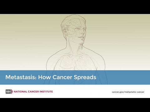
metastasis: how cancer spreads
Many cancer deaths are caused when cancer moves from the original tumor and spreads to other tissues and organs. This is called metastatic cancer. This animation shows how cancer cells travel from the place in the body where they first formed to other parts of the body.
Treatment for retinoblastoma depends on whether it is intraocular (within the eye) or extraocular (outside the eye).
Recurrent retinoblastoma is cancer that has recurred (come back) after it has been treated. The cancer may recur in the eye, in tissues around the eye, or in other places in the body.
Treatment Option Overview
KEY POINTS
There are different types of treatment for patients with retinoblastoma.
Children with retinoblastoma should have their treatment planned by a team of health care providers who are experts in treating cancer in children.
Treatment for retinoblastoma may cause side effects.
Six types of standard treatment are used:
Cryotherapy
Thermotherapy
Chemotherapy
Radiation therapy
High-dose chemotherapy with stem cell rescue
Surgery (enucleation)
New types of treatment are being tested in clinical trials.
Targeted therapy
Patients may want to think about taking part in a clinical trial.
Patients can enter clinical trials before, during, or after starting their cancer treatment.
Follow-up tests may be needed.
There are different types of treatment for patients with retinoblastoma.
Different types of treatment are available for patients with retinoblastoma. Some treatments are standard (the currently used treatment), and some are being tested in clinical trials. A treatment clinical trial is a research study meant to help improve current treatments or obtain information on new treatments for patients with cancer. When clinical trials show that a new treatment is better than the standard treatment, the new treatment may become the standard treatment.
Because cancer in children is rare, taking part in a clinical trial should be considered. Some clinical trials are open only to patients who have not started treatment.
Children with retinoblastoma should have their treatment planned by a team of health care providers who are experts in treating cancer in children.
The goals of treatment are to save the child's life, to save vision and the eye, and to prevent serious side effects. Treatment will be overseen by a pediatric oncologist, a doctor who specializes in treating children with cancer. The pediatric oncologist works with other health care providers who are experts in treating children with eye cancer and who specialize in certain areas of medicine. These may include a pediatricophthalmologist(children's eye doctor) who has a lot of experience in treating retinoblastoma and the following specialists:
Treatment for retinoblastoma may cause side effects.
For information about side effects that begin during treatment for cancer, see our Side Effects page.
Side effects from cancer treatment that begin after treatment and continue for months or years are called late effects. Late effects of treatment for retinoblastoma may include the following:
Physical problems such as seeing or hearing problems or, if the eye is removed, a change in the shape and size of the bone around the eye.
Changes in mood, feelings, thinking, learning, or memory.
Past treatment with radiation therapy, especially before age 1 year.
Having already had a previous second cancer.
It is important to talk with your child's doctors about the effects cancer treatment can have on your child. Regular follow-up by health professionals who are experts in diagnosing and treating late effects is important. See the PDQ summary on Late Effects of Treatment for Childhood Cancer for more information.
Six types of standard treatment are used:
Cryotherapy
Cryotherapy is a treatment that uses an instrument to freeze and destroy abnormaltissue. This type of treatment is also called cryosurgery.
Thermotherapy
Thermotherapy is the use of heat to destroy cancer cells. Thermotherapy may be given using a laser beam aimed through the dilatedpupil or onto the outside of the eyeball. Thermotherapy may be used alone for small tumors or combined with chemotherapy for larger tumors. This treatment is a type of laser therapy.
Chemotherapy
Chemotherapy is a cancer treatment that uses drugs to stop the growth of cancer cells, either by killing the cells or by stopping them from dividing. The way the chemotherapy is given depends on the stage of the cancer and where the cancer is in the body.
There are different types of chemotherapy:
Systemic chemotherapy: When chemotherapy is taken by mouth or injected into a veinor muscle, the drugs enter the bloodstream and can reach cancer cells throughout the body. Systemic chemotherapy is given to shrink the tumor (chemoreduction) and avoid surgery to remove the eye. After chemoreduction, other treatments may include radiation therapy, cryotherapy, laser therapy, or regional chemotherapy.
Systemic chemotherapy may also be given to kill any cancer cells that are left after the initial treatment or to patients with retinoblastoma that occurs outside the eye. Treatment given after the initial treatment, to lower the risk that the cancer will come back, is called adjuvant therapy.
Regional chemotherapy: When chemotherapy is placed directly into the cerebrospinal fluid (intrathecal chemotherapy), an organ (such as the eye), or a body cavity, the drugs mainly affect cancer cells in those areas. Several types of regional chemotherapy are used to treat retinoblastoma.
Ophthalmic artery infusion chemotherapy: Ophthalmic artery infusion chemotherapy carries anticancer drugs directly to the eye. A catheter is put into an artery that leads to the eye and the anticancer drug is given through the catheter. After the drug is given, a small balloon may be inserted into the artery to block it and keep most of the anticancer drug trapped near the tumor. This type of chemotherapy may be given as the initial treatment when the tumor is in the eye only or when the tumor has not responded to other types of treatment. Ophthalmic artery infusion chemotherapy is given at special retinoblastoma treatment centers.
Intravitreal chemotherapy: Intravitreal chemotherapy is the injection of anticancer drugs directly into the vitreous humor (jelly-like substance) inside of the eye. It is used to treat cancer that has spread to the vitreous humor and has not responded to treatment or has come back after treatment.
Radiation therapy is a cancer treatment that uses high-energy x-rays or other types of radiation to kill cancer cells or keep them from growing. There are two types of radiation therapy:
External-beam radiation therapy uses a machine outside the body to send radiation toward the cancer. Certain ways of giving radiation therapy can help keep radiation from damaging nearby healthy tissue. These types of radiation therapy include the following:
Intensity-modulated radiation therapy (IMRT): IMRT is a type of 3-dimensional (3-D) external radiation therapy that uses a computer to make pictures of the size and shape of the tumor. Thin beams of radiation of different intensities (strengths) are aimed at the tumor from many angles.
Proton-beam radiation therapy: Proton-beam therapy is a type of high-energy, external radiation therapy. A radiation therapy machine aims streams of protons(tiny, invisible, positively-charged particles) at the cancer cells to kill them.
Internal radiation therapy uses a radioactive substance sealed in needles, seeds, wires, or catheters that are placed directly into or near the cancer. Certain ways of giving radiation therapy can help keep radiation from damaging nearby healthy tissue. This type of internal radiation therapy may include the following:
Plaque radiotherapy: Radioactive seeds are attached to one side of a disk, called a plaque, and placed directly on the outside wall of the eye near the tumor. The side of the plaque with the seeds on it faces the eyeball, aiming radiation at the tumor. The plaque helps protect other nearby tissue from the radiation.ENLARGEPlaque radiotherapy of the eye. A type of radiation therapy used to treat eye tumors. Radioactive seeds are placed on one side of a thin piece of metal (usually gold) called a plaque. The plaque is sewn onto the outside wall of the eye. The seeds give off radiation which kills the cancer. The plaque is removed at the end of treatment, which usually lasts for several days.
The way the radiation therapy is given depends on the type and stage of the cancer being treated and how the cancer responded to other treatments. External and internal radiation therapy are used to treat retinoblastoma.
High-dose chemotherapy with stem cell rescue
High doses of chemotherapy are given to kill cancer cells. Healthy cells, including blood -forming cells, are also destroyed by the cancer treatment. Stem cell rescue is a treatment to replace the blood-forming cells. Stem cells (immature blood cells) are removed from the blood or bone marrow of the patient and are frozen and stored. After the patient completes chemotherapy, the stored stem cells are thawed and given back to the patient through an infusion. These reinfused stem cells grow into (and restore) the body's blood cells.
Enucleation is surgery to remove the eye and part of the optic nerve. A sample of the eye tissue that is removed will be checked under a microscope to see if there are any signs that the cancer is likely to spread to other parts of the body. This should be done by an experienced pathologist, who is familiar with retinoblastoma and other diseases of the eye. Enucleation is done if there is little or no chance that vision can be saved and when the tumor is large, did not respond to treatment, or comes back after treatment. The patient will be fitted for an artificial eye.
Close follow-up is needed for 2 years or more to check for signs of recurrence in the area around the affected eye and to check the other eye.
New types of treatment are being tested in clinical trials.
This summary section describes treatments that are being studied in clinical trials. It may not mention every new treatment being studied. Information about clinical trials is available from the NCI website.
Targeted therapy
Targeted therapy is a type of treatment that uses drugs or other substances to attack cancer cells. Targeted therapies usually cause less harm to normal cells than chemotherapy or radiation therapy do.
Targeted therapy is being studied for the treatment of retinoblastoma that has recurred(come back).
Patients may want to think about taking part in a clinical trial.
For some patients, taking part in a clinical trial may be the best treatment choice. Clinical trials are part of the cancer research process. Clinical trials are done to find out if new cancer treatments are safe and effective or better than the standard treatment.
Many of today's standard treatments for cancer are based on earlier clinical trials. Patients who take part in a clinical trial may receive the standard treatment or be among the first to receive a new treatment.
Patients who take part in clinical trials also help improve the way cancer will be treated in the future. Even when clinical trials do not lead to effective new treatments, they often answer important questions and help move research forward.
Patients can enter clinical trials before, during, or after starting their cancer treatment.
Some clinical trials only include patients who have not yet received treatment. Other trials test treatments for patients whose cancer has not gotten better. There are also clinical trials that test new ways to stop cancer from recurring (coming back) or reduce the side effects of cancer treatment.
Clinical trials are taking place in many parts of the country. Information about clinical trials supported by NCI can be found on NCI’s clinical trials search webpage. Clinical trials supported by other organizations can be found on the ClinicalTrials.gov website.
Follow-up tests may be needed.
Some of the tests that were done to diagnose the cancer or to find out the stage of the cancer may be repeated. Some tests will be repeated in order to see how well the treatment is working. Decisions about whether to continue, change, or stop treatment may be based on the results of these tests.
Some of the tests will continue to be done from time to time after treatment has ended. The results of these tests can show if your child's condition has changed or if the cancer has recurred (come back). These tests are sometimes called follow-up tests or check-ups.
If the tumor is large and it is not likely that the eye can be saved, treatment may include the following:
Surgery (enucleation). After surgery, systemic chemotherapy may be given to lower the risk that the cancer will spread to other parts of the body.
When retinoblastoma is in both eyes, the treatment for each eye may be different, depending on the size of the tumor and whether it is likely that the eye can be saved. The dose of systemic chemotherapy is usually based on the eye that has more cancer.
Treatment for cavitary retinoblastoma, a type of intraocular retinoblastoma, may include the following:
Systemic chemotherapy or ophthalmic artery infusion chemotherapy.
Use our clinical trial search to find NCI-supported cancer clinical trials that are accepting patients. You can search for trials based on the type of cancer, the age of the patient, and where the trials are being done. General information about clinical trials is also available.
Treatment of Extraocular Retinoblastoma
Treatment for extraocularretinoblastoma that has spread to the area around the eye may include the following:
It is not clear whether treatment with chemotherapy, radiation therapy, or high-dose chemotherapy with stem cell rescue helps patients with extraocular retinoblastoma live longer.
For trilateral retinoblastoma, treatment may include the following:
Systemic chemotherapy followed by high-dose chemotherapy with stem cell rescue.
Systemic chemotherapy followed by surgery and external-beam radiation therapy.
For retinoblastoma that has spread to other parts of the body, but not the brain, treatment may include the following:
Chemotherapy followed by high-dose chemotherapy with stem cell rescue and external-beam radiation therapy.
Use our clinical trial search to find NCI-supported cancer clinical trials that are accepting patients. You can search for trials based on the type of cancer, the age of the patient, and where the trials are being done. General information about clinical trials is also available.
Treatment of Progressive or Recurrent Retinoblastoma
A clinical trial that checks a sample of the patient's tumor for certain gene changes. The type of targeted therapy that will be given to the patient depends on the type of gene change.
Treatment of progressive or recurrent extraocular retinoblastoma may include the following:
Systemic chemotherapy and external-beam radiation therapy for retinoblastoma that comes back after surgery to remove the eye.
A clinical trial that checks a sample of the patient's tumor for certain gene changes. The type of targeted therapy that will be given to the patient depends on the type of gene change.
Use our clinical trial search to find NCI-supported cancer clinical trials that are accepting patients. You can search for trials based on the type of cancer, the age of the patient, and where the trials are being done. General information about clinical trials is also available.
To Learn More About Childhood Cancer
For more information from the National Cancer Institute about the treatment of retinoblastoma, see the following:
Physician Data Query (PDQ) is the National Cancer Institute's (NCI's) comprehensive cancer information database. The PDQ database contains summaries of the latest published information on cancer prevention, detection, genetics, treatment, supportive care, and complementary and alternative medicine. Most summaries come in two versions. The health professional versions have detailed information written in technical language. The patient versions are written in easy-to-understand, nontechnical language. Both versions have cancer information that is accurate and up to date and most versions are also available in Spanish.
PDQ is a service of the NCI. The NCI is part of the National Institutes of Health (NIH). NIH is the federal government’s center of biomedical research. The PDQ summaries are based on an independent review of the medical literature. They are not policy statements of the NCI or the NIH.
Purpose of This Summary
This PDQ cancer information summary has current information about the treatment of retinoblastoma. It is meant to inform and help patients, families, and caregivers. It does not give formal guidelines or recommendations for making decisions about health care.
Reviewers and Updates
Editorial Boards write the PDQ cancer information summaries and keep them up to date. These Boards are made up of experts in cancer treatment and other specialties related to cancer. The summaries are reviewed regularly and changes are made when there is new information. The date on each summary ("Updated") is the date of the most recent change.
The information in this patient summary was taken from the health professional version, which is reviewed regularly and updated as needed, by the PDQ Pediatric Treatment Editorial Board.
Clinical Trial Information
A clinical trial is a study to answer a scientific question, such as whether one treatment is better than another. Trials are based on past studies and what has been learned in the laboratory. Each trial answers certain scientific questions in order to find new and better ways to help cancer patients. During treatment clinical trials, information is collected about the effects of a new treatment and how well it works. If a clinical trial shows that a new treatment is better than one currently being used, the new treatment may become "standard." Patients may want to think about taking part in a clinical trial. Some clinical trials are open only to patients who have not started treatment.
Clinical trials can be found online at NCI's website. For more information, call the Cancer Information Service (CIS), NCI's contact center, at 1-800-4-CANCER (1-800-422-6237).
Permission to Use This Summary
PDQ is a registered trademark. The content of PDQ documents can be used freely as text. It cannot be identified as an NCI PDQ cancer information summary unless the whole summary is shown and it is updated regularly. However, a user would be allowed to write a sentence such as “NCI’s PDQ cancer information summary about breast cancer prevention states the risks in the following way: [include excerpt from the summary].”
Images in this summary are used with permission of the author(s), artist, and/or publisher for use in the PDQ summaries only. If you want to use an image from a PDQ summary and you are not using the whole summary, you must get permission from the owner. It cannot be given by the National Cancer Institute. Information about using the images in this summary, along with many other images related to cancer can be found in Visuals Online. Visuals Online is a collection of more than 3,000 scientific images.
Disclaimer
The information in these summaries should not be used to make decisions about insurance reimbursement. More information on insurance coverage is available on Cancer.gov on the Managing Cancer Care page.
Contact Us
More information about contacting us or receiving help with the Cancer.gov website can be found on our Contact Us for Help page. Questions can also be submitted to Cancer.gov through the website’s E-mail Us.
ver historia personal en: www.cerasale.com.ar [dado de baja por la Cancillería Argentina por temas políticos, propio de la censura que rige en nuestro medio]//
www.revistamedicos.com.ar //
www.quorumtuc.com.ar //
www.sectorsalud.com.ar //
www.maimonides.edu //
weblog.maimonides.edu/farmacia/archives/UM_Informe_Autoevaluacion_FyB.pdf - //
weblog.maimonides.edu/farmacia/archives/0216_Admin_FarmEcon.pdf - //
www.documentalistas.org.ar //
www.cpcesfe2.org.ar //
www.nogracias.eu //
www.estenssorome.com.ar //
www.cuautitlan.unam.mx/descargas/licenciaturas/bqd/plandestudio_bqd_ //
www.latamjpharm.org/trabajos/25/2/LAJOP_25_2_6_1_M4M6Z9746D.pdf //
www.nogracias.eu/v_juventud/informacion/informacionver.asp?cod= //
www.colfarse.com.ar //
www.proz.com/kudoz/english_to_spanish/art_literary/523942-key_factors.html - 65k - // www.llave.connmed.com.ar/portalnoticias_vernoticia.php?codigonoticia=17715 // www.frusculleda.com.ar/homepage/espanol/activities_teaching.htm // http://www.on24.com.ar/nota.aspx?idNot=36331 ||

 scan of the head and neck; drawing shows a child lying on a table that slides through the CT scanner, which takes x-ray pictures of the inside of the head and neck.")


.png)


No hay comentarios:
Publicar un comentario