Alzheimer’s screening may one day be carried out using an eye exam

A study conducted by researchers at Washington University School of Medicine suggests that it may be possible to use an eye test to screen people for their risk of developing Alzheimer’s disease.
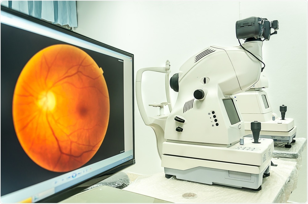 Image Credit: Arnon Thongkonghan / Shutterstock
Using a non-invasive eye exam, the researchers detected signs of Alzheimer’s in older people before they had developed any symptoms of the disease.
The plaques that occur in the brains of people with Alzheimer’s can start to accumulate up to twenty years before symptoms such as memory loss and cognitive decline develop.
O’Bryhim says the hope is that this new technique could be used to understand who is accumulating these abnormal proteins in the brain that may lead them to develop the disease.
Previously, studies have shown thinning in the retina and degradation of the optic nerve among patients who have died from Alzheimer’s.
"The retina and central nervous system are so interconnected that changes in the brain could be reflected in cells in the retina," said co-author Rajendra Apte.
For the study, the team used a non-invasive method called optical coherence tomography angiography to test the eyes of 30 individuals (average age in the mid-70s) who were not showing any symptoms of Alzheimer’s.
The test involves shining a light into the eye to assess retinal thickness and fiber thickness in the optic nerve.
One form of this test is already used in ophthalmologist’s offices, but in this study, the researchers added angiography to the test.
This angiography component enabled them to differentiate red blood cells from other retinal tissue to assess blood-flow patterns.
PET scans and lumbar punctures showed that 17 of the patients had high levels of amyloid or tau proteins, suggesting that they were at risk of developing Alzheimer’s.
As reported in the journal JAMA, those 17 patients also had thinning retinas and areas that lacked vasculature in the center of the retina. In the remainder of the patients, who had PET scans and lumbar puncture results that were within the normal range, retinas were healthy.
The researchers say more studies are needed to replicate the findings but that if the test can be used as a marker of Alzheimer’s risk, then it may be possible to screen people in their 40s or 50s to determine whether they are likely to develop the disease.
“If we could use this eye test to notice when the pathology is beginning, it may be possible one day to start treatments sooner to delay further damage,” says co-author Gregory Van Stavern.


.png)


No hay comentarios:
Publicar un comentario