Cool Videos: Patching and Sealing the Cell Membrane
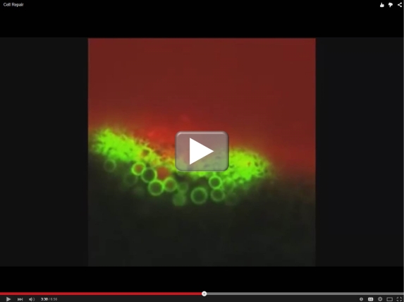
Bill Bement describes himself as a guy who “passionately, obsessively, and almost feverishly” loves to study cells. His excitement comes through in our final installment of the American Society for Cell Biology’s Celldance 2014. Bement, an NIH grantee at the University of Wisconsin, Madison, shares his scanning confocal microscope with us for this fascinating glimpse into the rapid response of cells to repair holes, tears, and other structural damage in their protective outer membranes.
For most people, this damage response runs on biochemical autopilot, sealing any membrane break within seconds to keep the cell viable and healthy. But some people inherit gene mutations that make sealing and patching difficult, particularly in cells that operate under repetitive mechanical stress. For example, some forms of muscular dystrophy stem specifically from an inherited inability to repair breaks in the cell membrane of skeletal muscle cells. In one type of disease that affects both skeletal and cardiac muscle, a gene mutation alters the shape of a protein called dysferlin, which normally binds annexin proteins that, as noted in the video, play a vital role in patching holes. In the presence of a glitch in dysferlin, the rapid chain of biochemical events needed to enable such repair breaks down.
There’s still an enormous amount to learn about cell membrane repair, so it will be interesting to see what Bement’s microscope and camera will show us next.
Links:
Bement Lab, University of Wisconsin-Madison
Celldance 2014, American Society for Cell Biology
NIH Support: National Institute of General Medical Sciences


.png)


No hay comentarios:
Publicar un comentario