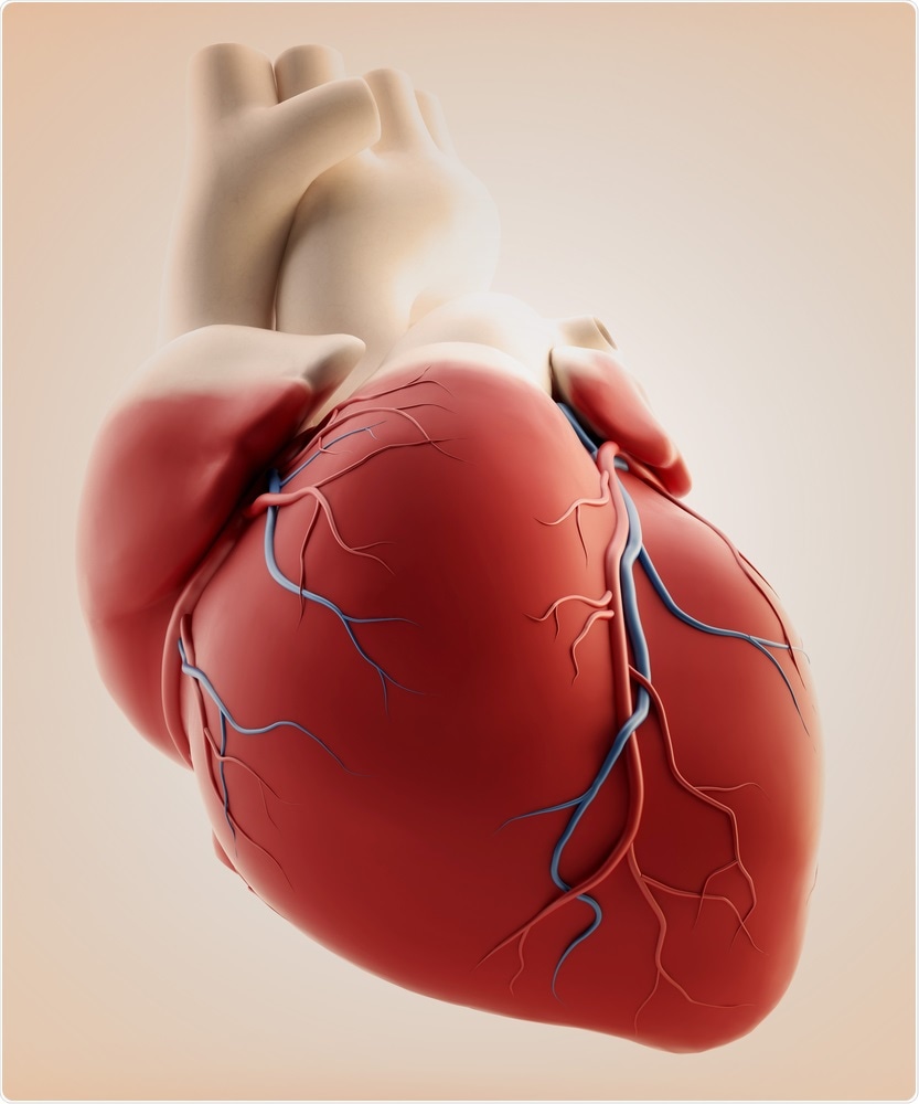
Defective immune signals could underlie some types of fatal heart disease

John Hopkins researchers have come up with exciting evidence that confirms the role of abnormal immune signaling in some types of inflammatory heart disease.
The research was recently published in the journal Cell Reports. In summary, it shows that among the two types of inflammatory cells called monocytes found in heart inflammation (myocarditis), the cytokine IL-17A inhibits the normal differentiation of the Ly6Clo type into macrophages.
This, in turn, promotes fibrous scarring, weakening, and dilation of the heart (called dilated cardiomyopathy or DCM), leading to eventual heart failure.
Myocarditis affects at least 1.5 million people a year and almost 10-16% of them develop DCM. There is no way at present to identify patients at risk of DCM. However, blocking IL-17A during myocarditis could effectively prevent DCM.
Previous research has shown that IL-17A is a cytokine produced in response to cardiac inflammation. It provokes the release of a fibroblast factor which disturbs the normal balance of two types of monocytes in the heart: Ly6Chi which promotes inflammation, and Ly6Clo which inhibits it.
An excessive number of Ly6Chi monocytes over Ly6Clo cells promotes DCM. Ly6Clo macrophages express more MHC II genes, which are associated with class II antigen processing. These cells help in resolving inflammatory damage and play an anti-inflammatory role.
Fibroblasts are collagen-producing cells in the body, normally responsible for mechanical and functional support of most organs in the body, and for wound repair.
In the heart, fibroblasts are crucial to forming and sustaining the form and function of the cardiac muscle or myocardium that acts as the heart pumps. It also forms fibrous tissue to replace dead cardiac myocytes, as following a heart attack.
In the current study, scientists found that fibroblasts also regulate the immune reaction during an episode of heart inflammation or myocarditis. This is disrupted by IL-17A to result in the fibrosis of the heart, a condition where the muscle cells are replaced by fibrous collagen. This reduces pumping efficiency and may lead to DCM.
How does IL-17A produce this effect?
To find out, the researchers joined the circulatory systems of two mice at a time to form several sets of conjoined mice. They then inoculated one mouse of each pair with cardiac myosin, a heart muscle protein, to produce experimental autoimmune myocarditis or EAM.
Once myocarditis set in, they examined macrophages from the hearts of both mice. They discovered that the macrophages in the inflamed heart came from both mice, which means that macrophages in myocarditis are drawn from the circulating pool rather than only those residing in the heart.
In the next step, they co-cultured Ly6Clo and Ly6Chi monocytes taken from the spleen of one mouse with fibroblasts extracted from the other mouse’s heart. They observed differentiation of almost all Ly6Chi monocytes into macrophages within 40 hours, but the differentiation of one-third of Ly6Clo cells took 160 hours.
Neither type differentiated or survived for long in the absence of cardiac fibroblasts. This shows the role played in monocyte differentiation by cytokines and other chemicals released by cardiac fibroblasts, regardless of their source.
They then cultured both types of monocytes in test tubes with cardiac fibroblasts, either unstimulated or after stimulating them with IL-17A, and found that in the second group, Ly6Clo monocytes remained completely undifferentiated after about a week, while the other type were unaffected.
This was repeated in live animals, with injections of each type of monocyte separately into the hearts of EAM-affected mice which are known to have IL-17A at high levels. The same pattern was confirmed.
In the IL-17A knockout mice, however, both types of monocytes underwent normal differentiation.
It is well known that IL-17A stimulated fibroblasts produce a protein called GM-CSF (granulocyte macrophage colony stimulating factor) that provokes inflammation. This molecule was found to be the mechanism by which IL-17A produces its effect. This was by mixing anti-GM-CSF antibodies with IL-17A, cardiac fibroblasts and Ly6Clo, which led to normal differentiation being restored.
Though IL-17A failed to prevent Ly6Chi monocyte differentiation, the Ly6Chi monocyte-derived macrophages showed lower levels of an enzyme called myeloid-epithelial-reproductive tyrosine kinase, or MerTK, produced in surface receptors. This enzyme is crucial to directing the phagocytosis of dead heart cells following injury or disease.
When it is too low, this process suffers, and toxins from dead cells cause delayed recovery from inflammation that may predispose to DCM. Self-antigens may be exposed which promote autoimmune damage to the heart muscle.
Human patients with myocarditis were found to have reduced MerTK levels than those with heart ischemia. This could mean the team has discovered a biomarker that could distinguish myocarditis from ischemia, two clinically similar conditions. However, more further research will be needed to confirm the study results.
Journal reference:
Hou X., et al., (2019). The Cardiac Microenvironment Instructs Divergent Monocyte Fates and Functions in Myocarditis. Cell Reports. https://doi.org/10.1016/j.celrep.2019.06.007


.png)


No hay comentarios:
Publicar un comentario