|
| | August 1, 2018 | |
| | |
| | The latest life science microscopy news from AZoNetwork | |
|
|
 |
| |  Using Synchronised Lasers for Time Resolved Microscopy Using Synchronised Lasers for Time Resolved Microscopy
Synchronising multiple ultrafast fiber lasers facilitates time-resolved microscopy at a high temporal resolution. This provides researchers the ability to observe dynamic cellular processes and discover new biological phenomena.
Find out more about developing such a laser system by reading the application note.
| |
|
|
|
|
 |
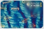
| |  Inspired by the beauty and breadth of images submitted for Image of the Year 2017, Olympus continues its quest for the best light microscopy art in 2018. For the chance to win a microscope or camera, applicants can now submit life science light microscopy images taken with Olympus equipment. Inspired by the beauty and breadth of images submitted for Image of the Year 2017, Olympus continues its quest for the best light microscopy art in 2018. For the chance to win a microscope or camera, applicants can now submit life science light microscopy images taken with Olympus equipment. | |
|
| |  Researchers at Cornell University have used ptychography to achieve the highest resolution ever produced in an electron microscope. The team, led by David Muller, Professor of Applied and Engineering Physics, recently reported their findings in the July 19th issue of Nature, outlining their ability to resolve images to a resolution of 0.39 å. Researchers at Cornell University have used ptychography to achieve the highest resolution ever produced in an electron microscope. The team, led by David Muller, Professor of Applied and Engineering Physics, recently reported their findings in the July 19th issue of Nature, outlining their ability to resolve images to a resolution of 0.39 å. | |
|
| |  Live cell imaging captures or visualizes human tissue in action. Several methods have been developed to study living cells in greater detail and with less effort, helping scientists gain a better grasp of biological functions. Live cell imaging captures or visualizes human tissue in action. Several methods have been developed to study living cells in greater detail and with less effort, helping scientists gain a better grasp of biological functions. | |
|
| |  ZEISS recently announced ZEISS ZEN Intellesis, a new machine learning capability that enables researchers to perform advanced analysis of their imaging samples across multiple microscopy methods. ZEISS recently announced ZEISS ZEN Intellesis, a new machine learning capability that enables researchers to perform advanced analysis of their imaging samples across multiple microscopy methods. | |
|
|
|
|


No hay comentarios:
Publicar un comentario