New probe allows identification of cancer stem cells in situ

A fluorescent molecular probe that targets cancer stem cells has been developed at the University of Illinois at Urbana-Champaign. The technology allows cancer stem cells to be studied in situ for the first time.
 Image Credit: Lightspring / Shutterstock
Cancer stem cells are the progenitor cells that develop into tumour cells. They are difficult to detect and may remain in the body after a primary tumour has been successfully treated or surgically removed.
These residual cancer stem cells, which have survived chemotherapy, often develop into new tumours causing metastases. Typically, such recurrence of a cancer will be in a form that is more aggressive than the primary tumour and also resistant to treatment.
Although the pivotal role of cancer stem cells in recurrent and metastatic cancers has been appreciated for some time, their elusive nature has made them difficult to study in situ.
In addition, cancer stem cells can be hard to distinguish from standard tumour cells with conventional imaging techniques.
Furthermore, the specific tissue microenvironments in which they live are complex and cannot be accurately mimicked in cell cultures. Consequently, it has not been possible to fully characterise this important cell type.
Researchers at the University of Illinois have this week published details of a molecular probe they have developed that allows cancer stem cells to be readily identified, tracked and studied in their native in vivo environment.
The probe, called AlDeSense, is specific for aldehyde dehydrogenase 1A1 (ALDH1A1), which is is highly elevated in cancer stem cells.
The probe fluoresces when it combines with its target enzyme, making the cancer stem cells clearly visible with flow cytometry and confocal imaging.
The probe has effectively identified cancer stem cells in cultures of multiple human cancer cell lines as well as in live mice.
AlDeSense therefore has the potential to provide a clinical imaging tool suitable for the medical evaluation and study of a broad range of cancer types.
Of particular interest was the ability of the probe to highlight the path of injected cancer stem cells as they spread through the bodies of mice and became tumours.
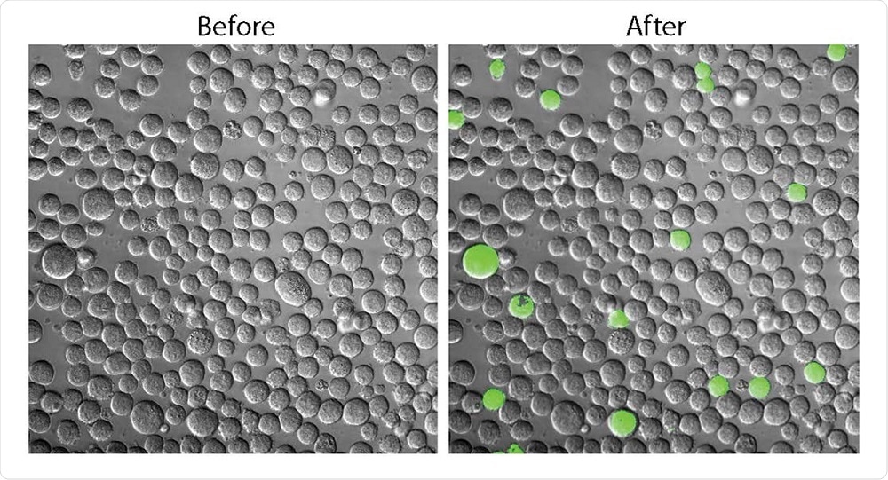 The A1DeSense probe makes it possible to visualise cancer stem cells (green). Image Credit: Chelsea Anorma, University of Illinois.
It is hoped that by making such imaging possible, AlDeSense will facilitate answering some fundamental questions regarding the behaviour of cancer stem cells.
It is hoped that the probe will also enable the prediction of cancer prognosis and facilitate screening for drugs that can kill cancer stem cells by targeting the ALDH1A1 enzyme.


.png)


No hay comentarios:
Publicar un comentario