Developing a Flexible X-ray Detector

 Thought LeadersDr. Imalka Jayawardena& Hashini ThirimanneThe University of Surrey
An interview with Dr. Imalka Jayawardena, PhD, and Ms Hashini Thirimanne, BSc, discussing the development of a flexible X-ray detector and how it can be applied to the diagnosis and treatment of human disease.
What are the problems faced by clinicians when analyzing and conducting X-rays?
Imalka: X-rays are a source of radiation that passes through the human body. Depending on the energy it has, some parts of it are absorbed at the tissue level and others penetrate further into the body.
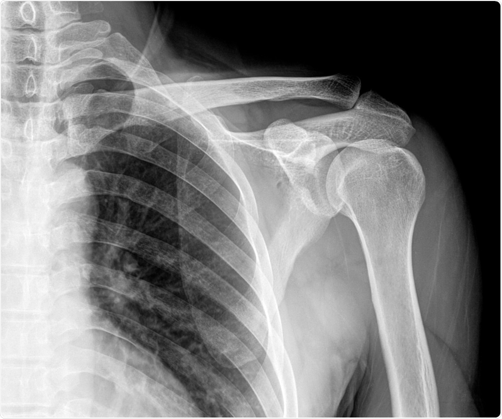 Image Credit: Xray Computer / Shutterstock
The problem is that the human body is not a flat surface and within the human body there are variations in tissue density that can affect whether X-rays are stopped and how they are scattered.
When you try to image the inside of the human body, scattering and changes in the stopping power of X-rays can distort the image, leading to errors in diagnosis, for example fractures or tumor sites, that could affect the patient treatment down the line.
There are also issues when it comes to treating patients using X-rays. In oncology, clinicians use X-rays to identify where the tumor lies and then use them to deliver specific doses of radiation in order to treat the patient.
The problem is because of the nature of X-rays the radiation spreads to surrounding areas other than the tumor location. That can cause secondary tissue damage, which can create the huge problem of new tumors.
Why is it important to develop new X-ray technologies?
Imalka: The modern way of imaging X-rays is based on what we call a flat panel architecture. This means the image made is really a flat structure, similar to the way the image is formed in a DSLR or mobile phone camera.
This means that at the moment we cannot image very complex shapes. When X-rays first started being used, old school film technology was used that allowed the bending of the film as needed into whatever shape was required, and then an image could be taken from that.
However, the disadvantage with this technology is that processing the film takes an hour, during which the patient cannot be diagnosed.
In certain cases, this time window can be quite critical, and so it is important to have a new generation of X-ray detector technologies where you can take a picture in real time, but at the same time image very complex shapes.
 Image Credit: Popartic / Shutterstock
Current technologies require very high X-ray doses. The higher the X-ray dose, the bigger the risk of causing further tissue damage and creating new tumors. Ideally we want to reduce the dose needed for imaging as well.
Overall, the development of new X-ray technologies that combine these three characteristics are needed: a low dose of X-rays, the ability to image very complex shapes, and the ability to do it in real time.
Please describe the X-ray detector you recently developed.
Hashini: Currently, the market comprises X-ray film technology and modern digital imaging X-rays. We recently released a model that combines these two, using a developed flexible X-ray detector which enables a real time response.
We have spent the last few years developing this flexible X-ray detector, which can be used as a personal radiation dosimeter. In simple terms, it is a flexible detector mounted onto an adhesive plaster and it can monitor the X-rays in an environment, powered by only a couple of wristwatch batteries.
It gives a response there and then - you do not need to have any specialized post-labs or anything to analyze it.
The technology is mainly based on a new material that we have developed. This material similar to an ink and because of this, it can be coated over very large areas and on any types of substrate.
This allows you to coat the ink on a plastic film, which is bendable, creating a flexible x-ray detector. In other words, the ink means that the technology is not constrained by size or flexibility.
How will this help clinicians when diagnosing patients?
Imalka: The first thing would be that the clinicians would be able to diagnose where to deliver the x-ray dose more accurately. This would be useful in areas like cancer therapy and bone fracture imaging.
Using this technology, clinicians would not have to carry out any complex mathematical manipulations to identify precisely where the fractures are. Effectively it is allowing a more accurate diagnosis.
In terms of patient therapy, it would provide a more accurate localized treatment, minimizing the subsequent development of tumors or tissue damage.
What other industries could this technology be applied to?
Hashini: X-ray detectors are not just limited to medical industry, they are also commonly used in non-destructive testing. They are also used in non-destructive testing, to detect any fractures or defects which could present in large structures.
For instance, if you have a fracture or a defect in an airplane (for aviation industries), or a shape (for building industries), X-rays provide a non-destructive way of detecting defects.
Alternatively, if you have a large building and you want to see a crack in a wall, you can use an X-ray detector and an X-ray source to screen it and identify any cracks without having to tear it down.
Imalka: In the aviation industry, up to 70% of non-destructive testing using X-rays is still based on X-ray film, as you can bend the film to image complex objects.
The one reason why the industry is hesitant to convert to using modern flat panel detectors is that you cannot image very complex shapes.
Effectively, having a detector or an image that can be bent to shape will modernize the way non-destructive testing is carried out in those industries.
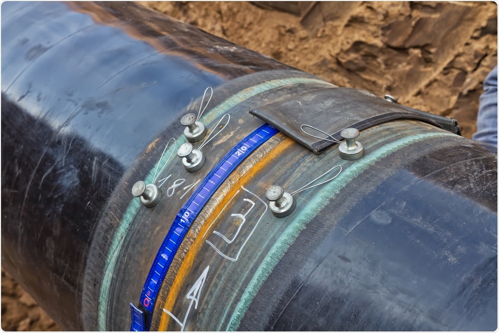 Non-destructive testing on a pipe. Image Credit: shinob / Shutterstock
Hashini: Another application for our technology would be in border security controls. For instance, in luggage screening if you had a large X-ray detector you could screen a higher volume or bigger luggage items in a very short time period.
The time constraint using our X-ray technology would be very low. Security applications would also be benefitted by our X-ray detectors as they can be portable due to them being powered by just a couple of batteries.
If there were suspicious objects lying around in a crowded place, the portable, flexible X-ray detectors would mean that the suspicious object could be covered up and screened instantly, with the result available there and then.
What are the next steps for your research?
Imalka: So far, we have developed a flexible X-ray detector and applied it to the first stage of X-ray imaging technology.
The X-ray imaging technology that we have at the moment is still based on a rigid system. It is still very much like the current camera sensors where everything is solid and you cannot manipulate it to the shape that you want.
What we are doing right now is working with industry to develop the back electronics that will help us to read out millions of pixels in one go whilst enabling flexibility.
That is the plan for the next five to ten years, and hopefully once we've optimized the whole array of detectors on those flexible plans, they will be brought into use in medical applications, non-destructive testing and in other fields like pharmaceuticals and food processing.
Where can readers find more information?
About Dr. Imalka Jayawardena
 Dr. Imalka Jayawardena is a postdoctoral researcher at the Advanced Technology Institute at the University of Surrey.
Imalka’s expertise covers a number of emerging optoelectronic systems, including organic and perovskite photovoltaics in addition to the organic-inorganic hybrid direct conversion X-ray detectors.
His work has resulted in over 30 peer reviewed publications and 2 invited conference presentations. He was also one of the winners for EPSRC PhD+ award made at the University if Surrey in 2012.
About Ms. Hashini Thirimanne
 Ms. Hashini Thirimanne is a Ph.D. student at the Advanced Technology Institute at the University of Surrey. She is currently working on “inorganics-in-organics” for high energy radiation detection.
Hashini was a finalist for the STEM for Britain 2018 competition and has been a part of a successful team that developed a first generation X-ray imager based on the organic-inorganic direct detector concept. She was also the Entrepreneurial support for the Large Area Pixelated X-ray Detector Project which was supported by the ICURe program.


.png)


No hay comentarios:
Publicar un comentario