Researchers discover how the heart becomes fully developed

Researchers from the Max Delbrück Center for Molecular Medicine (MDC) in Berlin have discovered how the heart develops from a simple tube into its characteristic S-shape and forms ventricles and atria.
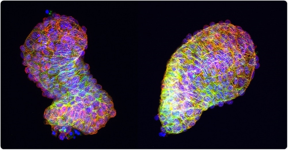 Image Credit: Anne M. Merks / MDC
The findings will help researchers improve their understanding of the development of congenital heart diseases.
As reported in Nature Communications, Daniela Panáková and colleagues identified the mechanism by which the heart takes on its fully developed form in zebrafish.
The study was published alongside another article in the same issue reporting on how the heart first develops into its simple tube-like form.
The researchers looked at how the linear tube loops round into the S-shape that ultimately goes on to form the ventricle and the atrium of the zebrafish heart.
Study author Anne Margarete Merks says that for this process to occur, the second-generation heart cells need to integrate into the linear heart and identify their correct place, which involves the cells relocating: "They change their neighbours and find new cells to share the cell boundaries with.”
The researchers say this process is controlled by a signalling pathway called the PCP (planar cell polarity) signaling pathway. Two particularly important components in this pathway are the molecules Fzd7a and Vangl2.
The PCP signaling pathway not only influences individual cells but affects the tissue as a whole. If this pathway is disrupted, the tissue tension changes and without the correct tension, the looping process does not take place and heart formation is impaired.
"Normally we observe that the cytoskeleton in the heart cells doesn't look the same everywhere, but exhibits polarity," explains Merks.
If the PCP signaling pathway is disrupted, this polarity is lost and the tube-shaped heart cannot take on its new form properly.
In particular, the outflow tract is unable to develop and problems with this part of the organ are the cause of most congenital heart diseases.
Merks says the team is confident that the findings can be transferred to mammals, including humans.
Next, Panáková and colleagues plan to carry out studies with heart tissue from patients with the congenital heart diseases.
They hope to identify exactly to what extent a disrupted PCP signalling pathway is implicated in the development of these diseases.


.png)


No hay comentarios:
Publicar un comentario