Economical Fluorescence Adapter for Stereo Microscopes

An interview with Charles Mazel, President of NIGHTSEA, conducted by Barbara Foster, The Microscopy & Imaging Place, Inc.
Before we get started - NIGHTSEA is an interesting name for a scientific instrument company. Where did that come from?
My fascination with fluorescence arose from SCUBA diving at night. A long time ago I asked myself “What happens if you dive at night with an ultraviolet light? Will anything fluoresce, and can you photograph it?” I eventually developed my own equipment to do this and the results were spectacular, especially on coral reefs. The resulting observations not only led to an abiding interest in fluorescence, both as a useful tool and for its inherent beauty, they changed my life.
I went on to earn a PhD in Marine Biology to try to understand what I was seeing. That led to a career in research, and then to invention and entrepreneurship. I was originally making equipment for other SCUBA divers, so the name NIGHTSEA, which I came up with before there was a company to attach it to, was very appropriate. The product line then grew into above-water laboratory applications to address varied needs of fluorescence observation.
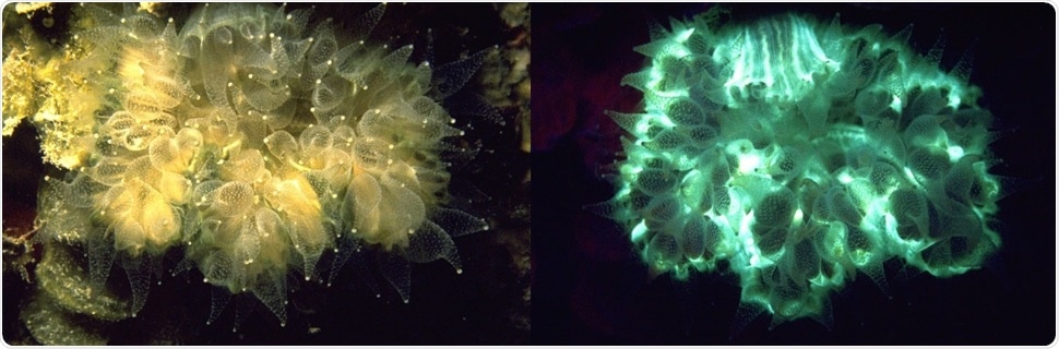
Coral photographed with white light (left) and fluorescing under ultraviolet light (right)
Why is fluorescence useful?
Not everything fluoresces, but when something does it can stand out brilliantly against its background, making it easy to see. In the natural world this makes fluorescence a powerful tool for finding subjects of interest, like coral polyps, In industry, fluorescence can highlight small defects or contaminants that might otherwise be hard to find. In forensic sciences fluorescence can be used to identify trace evidence, locate bodily fluids, and more. The Life Sciences use fluorescent proteins and dyes extensively to highlight structures and processes. As a matter of fact, the 2008 Nobel Prize in Chemistry was awarded for the discovery, development, and application of fluorescent proteins, especially for microscopy.
The benefits of fluorescent proteins and other fluorophores create an accompanying need for instrumentation to view and image them. The past 30 years have seen an explosion of fluorescence instrumentation, from the original widefield fluorescence microscope, to TIRF, FRET, confocal and two-photon microscopes, as well as super-resolution techniques.
More recently, fluorescence has moved into stereo microscopy, taking advantage of its lower magnification and three-dimensionality to view larger, bulkier objects like organs, embryos, and small animals. This is where our flagship product, the Model SFA Stereo Microscope Fluorescence Adapter, fits in. It lowers the barrier to add fluorescence to the suite of routine laboratory tools.
What has been the barrier to wider use of fluorescence in stereo microscopy?
A major stumbling block has been cost. Purpose-built fluorescence stereo microscopes can easily cost $20,000 and up. While they are wonderful research tools they are overkill for basic jobs like screening and sorting fluorescent zebrafish, Drosophila (fruit flies), frog (Xenopus) embryos, C. elegans, and seeds, as well as for checking that fluorescent staining is proper in a sample before moving on to higher-end imaging tools like confocal microscopes. An analogy I make is that it is like using a powerful word processing program to write a text message – you can use it, but you certainly don’t need it.
The SFA is a workhorse solution that addresses a wide range of fluorescence imaging challenges at an economical price. It’s shifting the whole fluorescence stereo microscopy paradigm. In comparison to a $20,000-30,000 research system, a complete setup to upgrade an existing stereo microscope with one excitation/emission wavelength combination is just under $1,100.
Because it is modular, you can add additional wavelength combinations at about half of that. There are thousands of unused stereo microscopes sitting in lab closets gathering dust. Our approach makes it practical to outfit those existing laboratory stereos with fluorescence, expanding science, education, industrial failure analysis. and more by lowering the barrier of entry for many users.
There is an additional benefit: the economical price tag also makes it practical to bring fluorescence into the classroom. It would be cost-prohibitive to provide multiple high-end fluorescence stereo microscopes in an undergraduate laboratory classroom, not to mention the wear-and-tear they would experience from untrained users. In comparison, robust, easy-to-use SFAs can upgrade 4, 6, or a dozen fluorescence stereos in the classroom, quickly, economically and with little risk of damage.
Can you describe the SFA system?
Fluorescence comes down to excitation and emission. You need to excite the fluorescing subject and then view it through an emission barrier filter that transmits the fluorescence while efficiently blocking the reflected excitation light. For excitation, we make interchangeable high intensity LED light heads that mount on the end of a flexible gooseneck lamp base. We currently offer five excitation wavelength options, plus a white light head. For each excitation source we have matching barrier filters and filter shields that mount on the microscope.
The barrier filter is positioned in the optical path below the objectives, while the filter shield projects out at a 45° angle. In addition to shielding the user from reflected excitation light the shield is also a barrier filter through which the fluorescence of the subject can be viewed directly. It’s always a delight to watch a newcomer to fluorescence view an object in normal “white light” then shift to see the same object through the filter shield. Even before looking in the microscope, they will often move back and forth between the normal view and the view through the filter, as if to really confirm that what they are seeing is real.
The only piece of the SFA system that touches the microscope is our adapter ring. It mounts with thumbscrews where a ring light would ordinarily attach. This makes it nearly universal, compatible with nearly every make and model of stereo microscope of any vintage. We have added alternative adapters for some of the outliers that the standard adapter doesn’t fit. We have also engineered custom solutions on a case-by-case basis for users with special requirements.
The system is fully modular. The light head attaches to the gooseneck with a bayonet connector. The barrier filter attaches to the adapter ring magnetically. The filter shield pivots into place on the adapter and is secured with a twist of a thumbscrew. You don’t need to acquire all the wavelength sets at the same time - buy what you need to get going and add additional wavelength combinations at any time. Need to change wavelengths? Just change the light head/barrier filter/shield combo. It only takes a matter of seconds. This video, “Zero to fluorescence in 40 seconds,” demonstrates just how easy it really is:
Did you originally develop the SFA system to address a specific market?
I never set out to develop a general-purpose fluorescence adapter. The prototype system that eventually became the SFA arose out of a project to develop fluorescence-based tools for studying coral recruitment, the process of baby corals (~1mm diameter) settling and growing on the reef. Their small size makes them essentially impossible to find under natural illumination conditions on complex reef surfaces. Fortunately, most corals are fluorescent and much of the rest of the reef is not, so you can take advantage of the power of fluorescence as a search tool.
Researchers often use artificial settlement tiles, essentially the rough sides of bathroom tiles, as surrogates for the natural reef. They place the tiles at different locations, depths, and orientations, then collect them months later and examine them under a stereo microscope to locate and identify corals. Part of our project was to determine if fluorescence was superior to the usual white light technique.
The answer was an unequivocal “Yes!” It was not a complete substitute for white-light viewing since not all corals fluoresce strongly, but it definitely helped in finding more corals faster, and finding many corals that could easily be missed under white light. But this was one of those times when the answer is ‘Yes, but so what?’ The value of fluorescence was clear, but fluorescence stereo microscopes were too expensive for most marine biology budgets. Also, it is not ideal to ship an expensive and delicate system back and forth to remote tropical islands.
.jpg?wmode=opaque)
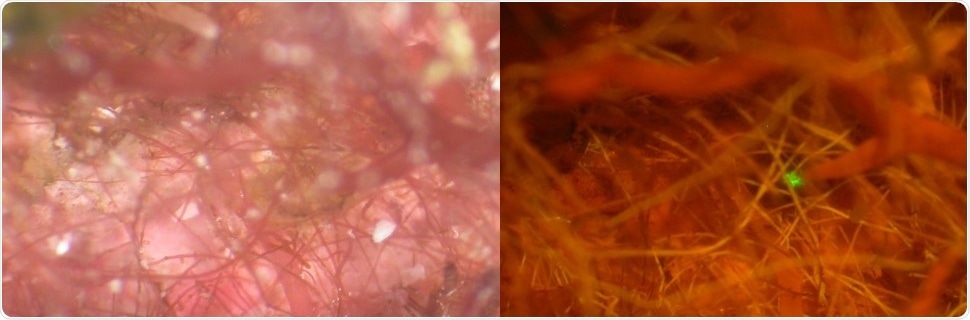
Stereo microscope images of a coral hidden in a ‘forest’ of algae under white light illumination (left) and in fluorescence. Photo credit Dr. Alina Szmant
To answer these challenges, I developed a prototype system for mounting high intensity LEDs and matching barrier filter on a conventional stereo microscope. The original system offered just one excitation/emission wavelength combination: blue light with a yellow longpass barrier filter that replicated the solution we had developed for underwater searching. While this microscope adapter functioned well, the project went no further at that time.
A few years later I was contacted by a researcher who needed to sort a large number of GFP-transgenic Drosophila. At that time we supplied a fluorescence excitation flashlight and filter glasses for working with larger subjects, and he had jury-rigged these to work with his microscope. He was really enthusiastic about the results. He presented such a compelling case for the Drosophila community needing an inexpensive solution for sorting that I created a new version of the microscope adapter and took it to their annual research conference. While the new adapter received a great reception, the researchers told us that they needed more than one wavelength combination, so it was back to the drawing board. I returned the next year with a modular two-wavelength system and the NIGHTSEA SFA was off and running!
How does the SFA compare to a high-end fluorescence stereo microscope? Why should someone consider getting the SFA instead of a purpose-built system?
The SFA can’t do everything that a high-end system can. If you have very small subjects, weak fluorescence, or need very high-resolution imaging you may really need a full research level system. However, for many tasks the SFA works just fine. As a rule of thumb, if a subject is easy to see on a high-end system there is a very good likelihood that it will be fine with the SFA. If you are having trouble seeing the fluorescence on an expensive system then the SFA is likely not the answer.
In practicality, the key metric is ‘Does it get the job done?’ Based on the feedback from our many users we know that the SFA is successful for a wide range of applications. In some cases it is the only fluorescence system a user has. In others it is taking the pressure off a high-end system so that it is not tied up with routine tasks like sorting. Also, researchers are using the system to put fluorescence where they would ordinarily not have it – in their zebrafish facility for screening transgenics without having to walk to another building; in their mouse facility for checking expression and aiding in dissection of fluorescent organs; and many more.
For labs that just can’t afford a purpose-built system, the SFA provides an important first step into stereo fluorescence microscopy. New faculty start-ups with limited budgets are a great example. And as mentioned above, the SFA now makes it affordable to include fluorescence in undergraduate teaching laboratories.
Is the system having an impact?
The SFA is helping people do their science. I recently had a new PhD approach me at a conference and tell me that the system had been critical in getting his thesis research done. NIGHTSEA equipment has been cited in the Methods section of more than 180 peer reviewed papers, and what we are especially proud of is that these appeared in more than 100 different journals, covering a vast array of disciplines. There is now NIGHTSEA equipment in more than 1300 research and educational institutions, in over 50 countries.
What applications are there beyond research?
We have already mentioned the use in undergraduate teaching laboratories. The SFA system is also being used for outreach. Its portability and ease of use make it practical to bring fluorescence microscopy along to middle schools, science fairs, mobile science labs, and the like.
Are there accessories or enhancements available for the system?
We have continued to enhance to the system based on customer feedback and requests. For example, we have expanded our product line from the original two excitation/emission combinations to the current six. We created a way for users to increase intensity by running two light heads simultaneously, with a custom mounting system that attaches to our adapter ring. We then took it a step further, adding a switch box to make it easy to alternate between two different excitation wavelengths.
Because of the ease of adding the SFA to just about any stereo microscope, customers were using it with microscopes that were not in a dark room. Sometimes that was fine, but in other situations light from overhead fixtures or nearby windows reduced the viewing contrast, making subjects hard to see. In response, we invented a novel ‘Tent of Darkness’ to surround the microscope. It provides near-complete darkness without sacrificing the ability to work with specimens on the sample stage. In combination with our battery pack that can run the system for more than eight hours you can literally explore the world of fluorescence anywhere, at any time.
We have more things in the pipeline, all based on customer requests.
Where can readers find more information?
The SFA page on the NIGHTSEA website.
About Dr. Charles Mazel
 Charles Mazel is the Founder and President of NIGHTSEA. Prior to founding NIGHTSEA and BlueLine NDT he was a Principal Research Scientist at Physical Sciences Inc. where he was involved in diverse research projects primarily related to underwater optical properties, including development of a novel diver-operated underwater fluorescence/reflectance spectrometer.
Charles’ earlier work history includes: deep ocean hydrographic and oceanographic survey work with Western Electric Corp.; field and training engineer for side scan sonars and sub-bottom profilers at Klein Associates Inc.; Research Engineer at MIT, managing the Hydrodynamics Laboratory; Assistant Director of the Edgerton Center at MIT, including teaching the strobe project laboratory course.
Charles Mazel has a Bachelor’s in Physics from Brandeis University, a Masters in Ocean Engineering from MIT, and a PhD in Biology from Boston University.
Sponsored Content Policy: News-Medical.net publishes articles and related content that may be derived from sources where we have existing commercial relationships, provided such content adds value to the core editorial ethos of News-Medical.Net which is to educate and inform site visitors interested in medical research, science, medical devices and treatments.


.png)


No hay comentarios:
Publicar un comentario