Creative Minds: Breaking Size Barriers in Cryo-Electron Microscopy

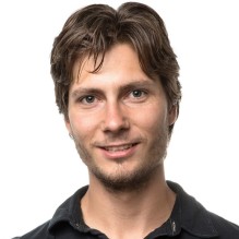
Dmitry Lyumkis
When Dmitry Lyumkis headed off to graduate school at The Scripps Research Institute, La Jolla, CA, he had thoughts of becoming a synthetic chemist. But he soon found his calling in a nearby lab that imaged proteins using a technique known as single-particle cryo-electron microscopy (EM). Lyumkis was amazed that the team could take a purified protein, flash-freeze it in liquid nitrogen, and then fire electrons at the protein, capturing the resulting image with a special camera. Also amazing was the sophisticated computer software that analyzed the raw 2D camera images, merging the data and reconstructing it into 3D representations of the protein.
The work was profoundly complex, but Lyumkis thrives on solving extremely difficult puzzles. He joined the Scripps lab to become a structural biologist and a few years later used single-particle cryo-EM to help determine the atomic structure of a key protein on the surface of the human immunodeficiency virus (HIV), the cause of AIDS. The protein had been considered one of the greatest challenges in structural biology and a critical target in developing an AIDS vaccine [1].
Now, Lyumkis has plans to take single-particle cryo-EM to a whole new level—literally. He wants to develop new methods that allow it to model the atomic structures of much smaller proteins. Right now, single-particle cryo-EM has worked with proteins as small as roughly 150 kilodaltons, a measure of a protein’s molecular weight (the approximate average mass of a protein is 53 kDa). Lyumkis plans to drop that number well below 100 kDa, noting that if his new methods work as he hopes, there should be very little, if any, lower size limit to get the technique to work. He envisions generating within a matter of days or weeks the precise structure of an average-sized protein involved in a disease, and then potentially handing it off as an atomic model for drug developers to target for more effective treatment.
To help him shrink the size barrier in cryo-EM, Lyumkis received a 2015 NIH Director’s Early Independence Award. Lyumkis, now at the Salk Institute for Biological Studies, La Jolla, has launched his own lab and is busy working out methods to solve a specific technical problem in cryo-EM. Currently, proteins less than 100 kDa don’t generate enough of a signal during imaging for existing computational tools to crunch through the raw 2D data. What happens is the visual background noise inherent to cryo-EM dominates the picture, making it extremely difficult to detect and analyze small proteins.
Lyumkis proposes to boost the signal a bit more by anchoring the proteins to larger macromolecular structures that are well characterized and relatively straightforward to use, such as ribosomes. With the protein anchored, he and colleagues can also develop algorithms that computationally exclude the extraneous regions of the image outside the protein of interest. This will allow researchers to zoom in on the signal and generate the needed data to reconstruct the atomic structures of these smaller proteins.
One of his test cases involves the multi-protein IKK complex, the master regulator of the NF-kB cellular pathway that is dysregulated in many cancers and a range of inflammatory conditions. The complex, much like the HIV protein that Lyumkis previously helped to solve, has presented many challenges to structural biologists since its discovery nearly 20 years ago. Lyumkis plans to demonstrate the improved size capabilities of single-particle cryo-EM by reconstructing smaller IKK protein domains that might elude his ongoing efforts to understand the complex as a whole.
If Lyumkis succeeds in shrinking the size barrier, it will be another important step forward for this 40-year-old technique that has gotten dramatically better in the last few years. In fact, the resolution of single-particle cryo-EM has improved so fast and so much, the journal Nature Methodsnamed cryo-EM as the Method of the Year in 2015. These improvements have major implications for drug discovery and development. It’s also why the work in the Lyumkis lab will be interesting to follow in the years ahead.
Reference:
[1] Cryo-EM structure of a fully glycosylated soluble cleaved HIV-1 envelope trimer. Lyumkis D, Julien JP, de Val N, Cupo A, Potter CS, Klasse PJ, Burton DR, Sanders RW, Moore JP, Carragher B, Wilson IA, Ward AB. Science. 2013 Dec 20;342(6165):1484-1490.
Links:
Lyumkis Lab (The Salk Institute for Biological Studies, La Jolla, CA)
Video: Dmitry Lyumkis: Electron Microscopy (The Salk Institute)
Lyumkis NIH Project Information (NIH RePORTER)
NIH Director’s Early Independence Award Program (Common Fund)
NIH Support: Common Fund; National Institute of Dental and Craniofacial Research


.png)


No hay comentarios:
Publicar un comentario