|
| | October 23, 2019 | |
| | Life Science Microscopy | |
| | The latest life science microscopy news from News Medical | |
|
|
 |
| |  Alignment of the Movable Objective Microscope Alignment of the Movable Objective Microscope
Two-photon imaging systems rely on accurate beam alignment for optimal performance. In this tutorial, we will walk through the steps of aligning a Sutter two-photon microscope using a special laser alignment tool and custom pinholes for the Sutter Movable Objective Microscope (MOM®).
| |
|
|
|
|
|
 |
| | Super-Resolution Microscopy vs. Electron Microscopy 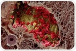 Conventional light microscopy has its limits. Light, being a wave, is subject to diffraction which severely limits the size of structures one can resolve. To be able to observe parts of an organism smaller than this, other techniques need to be employed. Conventional light microscopy has its limits. Light, being a wave, is subject to diffraction which severely limits the size of structures one can resolve. To be able to observe parts of an organism smaller than this, other techniques need to be employed. | |
|
| | Confocal Reflection Microscopy for Cell Migration Studies  Cell migration occurs throughout life as a response to internal and external signals, and can as such be indicative of changes occurring in the body. Imaging this movement and what processes control it can be critical in understanding cell behavior, development, and disease. Cell migration occurs throughout life as a response to internal and external signals, and can as such be indicative of changes occurring in the body. Imaging this movement and what processes control it can be critical in understanding cell behavior, development, and disease. | |
|
| | An Introduction to IR Nanochemical Mapping  AFM-IR compositional mapping of Streptomyces bacteria. Left: AFM topographic image of bacterial cells. Middle: AFM-IR absorption at 1650 cm−1, corresponding to the amide I band associated with protein. Right: AFM-IR absorption at the carbonyl band 1740 cm−1, indicating the distribution of triglyceride vesicles within bacterial cells. AFM-IR compositional mapping of Streptomyces bacteria. Left: AFM topographic image of bacterial cells. Middle: AFM-IR absorption at 1650 cm−1, corresponding to the amide I band associated with protein. Right: AFM-IR absorption at the carbonyl band 1740 cm−1, indicating the distribution of triglyceride vesicles within bacterial cells. | |
|
|
|
|


.png)


No hay comentarios:
Publicar un comentario