How is Takayasu’s Arteritis Diagnosed?
Skip to:
- What is Takayasu's arteritis?
- What causes Takayasu's arteritis and who is at risk?
- Management and diagnosis of Takayasu's arteritis using non-invasive imaging
- Summary
What is Takayasu's arteritis?
Vasculitis is an inflammation of blood vessels. Takayasu's arteritis is one form of vasculitis, in which chronic inflammation affects the walls of the aorta and other large arteries. In later stages the disease leads to thickening of vessel walls, mural edema and mural enhancement. The function of the aorta is to supply blood to the head, arms, internal organs and legs. The inflammation leads to the thickening of the walls with resulting stenosis, or narrowing, of the blood vessel.
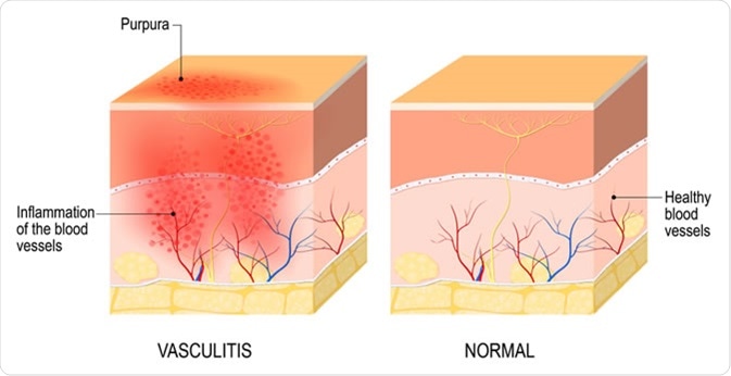
Vasculitis, damange of blood vessels by inflammation. Designua / Shutterstock
This narrowing of the blood vessels could lead to a reduced blood flood hence less oxygen is transported to the body parts and organs supplied by the artery.
Symptoms of stenosis can include dizziness, fainting or headaches, chest pain, stroke, heart attack as well as pain when arms and legs are used. During stenosis, smaller vessels grow to allow the transport of blood around the blockage. These vessels are known as collateral vessels, which could help to prevent organ damage.
Another symptom of Takayasu's arteritis includes fevers, a blood pressure difference in both arms caused by stenosis and change in color of legs and hands due to a restricted blood flow. However, these symptoms are not specific and therefore diagnosis could be delayed.
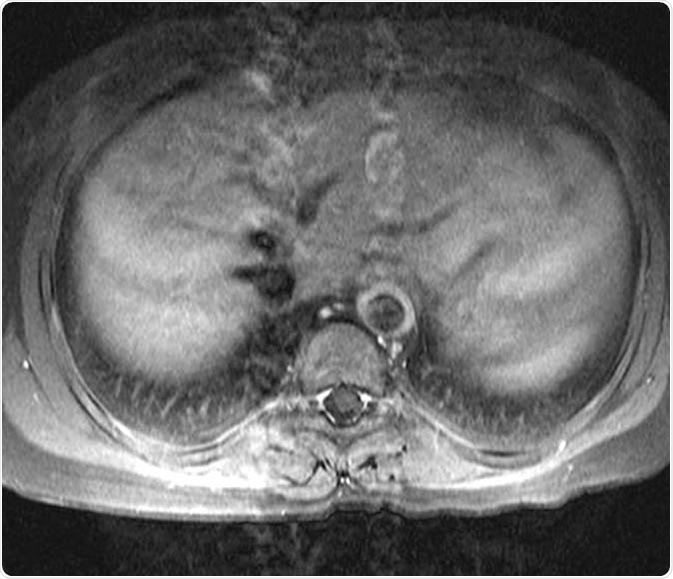
15 year-old female patient with known Takayasu arteritis presented for MRI to investigate back pain. The axial T1-weighted post-gadolinium MRI above shows thickened, enhancing aortic wall, consistent with large vessel vasculitis. Dr Laughlin Dawes, http://www.radpod.org/2007/04/15/takayasu-arteritis/
What causes Takayasu's arteritis and who is at risk?
The causes of Takayasu's arteritis are not known. It is classed as an autoimmune disease, which means the immune systems of the body attacks the blood vessels. Only one in around 200,000 people is affected, mostly at the age of 15-40. Some studies reported that mostly females from East Asia are affected. However, it has now been established that the disease can happen in any country.
Management and diagnosis of Takayasu's arteritis using non-invasive imaging
X-ray angiography or arteriography is the most common procedure for the diagnosis of Takayasu's arteritis. This procedure produces x-ray images of the blood vessels. However, early stages of the disease which could potentially be reversible can sometimes not be detected with this method.
Two non-invasive methods have been investigation to evaluate their ability in the early diagnosis of Takayasu's arteritis, magnetic resonance imaging (MRI) and fluorodeoxyglucose positron emission tomography (FDG-PET). With FDG-PET, it is possible to detect high glucose metabolic areas, which are typically seen when inflammation in tissues are present.
MRI produces high resolution images that show features of vascular inflammation. The thickening of vessel walls takes place very early in the inflammatory stage of the disease, which can be detected by MRI. This shows how important MRI is in the detection of early disease stages, when damage is more likely to be reversible.
Previously, intraoperative angiography was the main invasive method for the diagnosis and management of Takayasu´s arteritis. However, it requires a radiographic dye which could impair renal function and therefore it is only used to detect the change of lumen diameter, which occurs at the later stages of the disease.
In a study by Andrews et al. in 2004, it was suggested that the non-invasive methods MRI and FDG-PET can disclose important information about the disease such as the presence of vascular inflammation or the wall thickening compared to the common angiography. Additionally, FDG-PET can identify all inflamed vessel and could be more sensitive measuring continuing blood vessel inflammation compared to conventional methods.
During the study a reversible anatomical change of the vessel wall could be detected in one patient after treatment. Therefore, these methods are not only for the early diagnosis of Takayasu´s arteritis but also the assessment of the effects after a treatment. The study concluded that these non-invasive methods can help to diagnose the disease early on as well as assess response to treatment when compared to other assessment methods or invasive angiography. MRI and FDG-PET could be the future of Takayasu´s arteritis diagnosis and replace invasive angiography in many patients.
In a study by Kumar et al. in 1997, the sensitivity of MR angiography was reported at 91%, accuracy at 86% and a specificity of 81% for detection of aortic arch and aortic arch branch vessel lesions. Furthermore, these figures were similar to the detection of determining the severity of the lesion compared to angiography. Even for patients with prior false negative results, MRA allowed an accurate diagnosis of Takayasu´s arteritis in all patients during the study.
Summary
MRA and FDG-PET have been demonstrated as accurate methods for the diagnosis of early stages of Takayasu´s arteritis as well the ongoing assessment after treatment. As they are non-invasive, risks remain low compared to invasive methods. However, further studies are necessary to evaluate the future use of these methods and eventually replace existing angiography.
Sources
- Kerr G.S. 1995. Takayasu's arteritis. Rheumatic Diseases Clinics of North America. https://europepmc.org/abstract/med/8592736.
- Huston K. 2019. Takayasu's Arteritis. American college of rheumatology. https://www.rheumatology.org/I-Am-A/Patient-Caregiver/Diseases-Conditions/Takayasus-Arteritis.
- Andrews J. 2004. Non-invasive imaging in the diagnosis and management of Takayasu's arteritis. https://www.ncbi.nlm.nih.gov/pmc/articles/PMC1755083/.
- Kumar S. et al. 1997. Takayasu's arteritis: evaluation with three-dimensional time-of-flight MR angiography. European Radiology. https://doi.org/10.1007/s003300050107.
Further Reading
Last Updated: Oct 9, 2019


.png)


No hay comentarios:
Publicar un comentario