Cell-Matrix Adhesion Overview
Tissue structures are determined by both cell-cell adhesion and cell-matrix adhesion, with the latter describing interactions between cells and the extracellular matrix (ECM). The ECM is the collection of collagen fibers, proteoglycans and multi-adhesive matrix proteins that are secreted by cells to form an essential support structure.

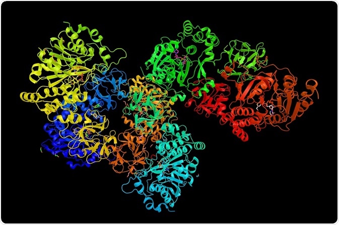
Credit: ibreakstock/ Shutterstock.com
Other functions of the ECM include segregating tissues and controlling communication between cells. Cell-matrix adhesion is formed through the utilization of cell adhesion molecules that bind to the cell surface of the ECM. Anchor proteins are also used to mediate linkages, or are employed in roles related to regulation.
The importance of integrins to cell-matrix adhesion
Integrins are a type of cell adhesion heterodimer molecule made up of two subunits that facilitate attachments between the cell surface and the ECM. The adhesion is produced via ligand binding sites composed from parts of both subunit chains.
Cells often express various types of integrins allowing for binding to multiple ECM molecules. This is important because integrins display weak interactions to their ligands and so large amounts of integrin to ECG protein binding sites are required for the cell to be firmly fixed to the matrix.
Furthermore, an advantage to this mechanism of multiple weak interactions between the cell and matrix is that it is necessary for migrating cells. The ability to quickly bind and separate from the matrix is essential when movement is required and the low affinity to ligands enables this.
Focal adhesions and hemidesmosomes as integrin-dependent junctions for cell-matrix adhesion
There are two types of integrin-dependent junctions where cell-matrix adhesion occurs. The first are called focal adhesions and are where the cytoskeleton of the cell attaches to the fibronectin glycoprotein of the ECM. Focal adhesions not only anchor the cell but also facilitate mechanical and biochemical signaling across the plasma membrane.
Also, integrin-cytoskeletal linkages at the focal adhesion can regulate cell behavior. For example, filamin is a component that connects the integrin chains to the actin cytoskeleton and inhibits cell migration as well as allowing mechanical forces to be sensed.
Hemidesmosomes are the second type of integrin-dependent junction and connect intermediate filaments to the basal laminae of epithelial cells. They are comprised of an inner and an outer plaque. The outer plaque is denser than the inner plaque and is directly connected to the inner surface of the membrane, with the inner plaque sandwiched in the middle.
The side of the plaque closest to the cytosol is composed of adapter proteins attached to keratin filament ends. Within hemisdesmosomes, integrin is localized and binds to the protein adaptor plectin and the ECM protein laminin. This type of integrin dependent junction improves the rigidity of epithelial tissues as found in the skin and corneas.
The influence of integrins on cell behavior
Integrins influence cell behavior by both providing an attachment site for the ECM and through the lateral connections with other proteins at the cell surface. The latter occurs through extracellular integrin receptor domains.
They can form multiple complexes with molecules employed for cell signaling and adaptor proteins connecting to the cytoskeleton of the cell. The cell adhesion to the ECM formed through integrin produces a bi-directional pathway for mechanical and biochemical information.
Intracellular signaling transmitting information in an 'outside-in' direction is important for cell spreading and cell migration. The opposite transference of information is an 'inside-out' direction which activates the ligand binding function of integrins.
Cross-talk between the two information directions has been identified suggesting a regulatory role and as a factor affecting integrin activation. As a consequence, cross-talk promotes 'outside-in' signaling through increasing intracellular or inside signals.
Reviewed by: Dr Tomislav Meštrović, MD, PhD
Sources:
- https://www.ncbi.nlm.nih.gov/books/NBK21539/
- https://www.ncbi.nlm.nih.gov/pubmed/17680633
- http://www.sciencedirect.com/science/article/pii/S0167488904000990
- http://www.sciencedirect.com/science/article/pii/S0022202X15404427
- https://www.ncbi.nlm.nih.gov/pmc/articles/PMC3479359/


.png)


No hay comentarios:
Publicar un comentario