
A High Resolution Look into Winemaking and Wine Growing
Words by Dr. Cameron Chai
Images by TESCAN
Images by TESCAN
Microscopy is a vital tool in the arsenal of life science researchers. There are a vast array of different microscopy techniques that enable researchers to see features and functionalities that are typically impossible to see with the naked eye. Similarly, life science covers a broad range of disciplines from cell biology and virology, through to etymology and agriculture.
Electron microscopy including the techniques Scanning Electron Microscopy (SEM) and Transmission Electron Microscopy (TEM) are imaging modalities that enable researchers to image features down to the nano and atomic scale. They go way beyond the capabilities of more common light microscopes. The region around Brno in the Czech Republic is credited as being the European cradle of electron microscopy.
Brno also happen to be the capital of the Moravian region in the south of the Czech Republic that borders Austria and Slovakia. This is the nation’s main wine producing region, responsible for 96% of their total capacity. While most wine from the region is consumed domestically, privatisation and recent changes to viticulture laws have lead to increasing exports to Europe, the US and Asia, following a period of isolation after the Second World War.
The geographical commonality lead to a collaborative project between TESCAN, a local manufacturer of SEMs and the National Wine Centre in the Czech Republic. This project saw TESCAN produce a series of intriguing images from samples provided by the Department of Viticulture and Viniculture at Mendel University, also located in Brno.
While there is no doubt that oenology, or winemaking is a science, it is rare that it collides with the cutting edge of science as it has here. The images produced in this project have no doubt provided a more detailed insight into winemaking and viticulture (wine growing) for those intimately involved in it, and for those who have benefitted from it!
These images formed part of a public display hosted by the National Wine Centre, at their headquarters in the Valtice Castle, part of a UNESCO world heritage site. Pavel Krška, Director of the National Wine Centre, said, “As far as we know, this exhibition is the first of its type and the feedback from visitors has been extremely positive. They are in awe of the images and visitors appreciate the fact that they are being given access to highly scientific content presented in such a way that it appeals to wine aficionados and the broader community.”
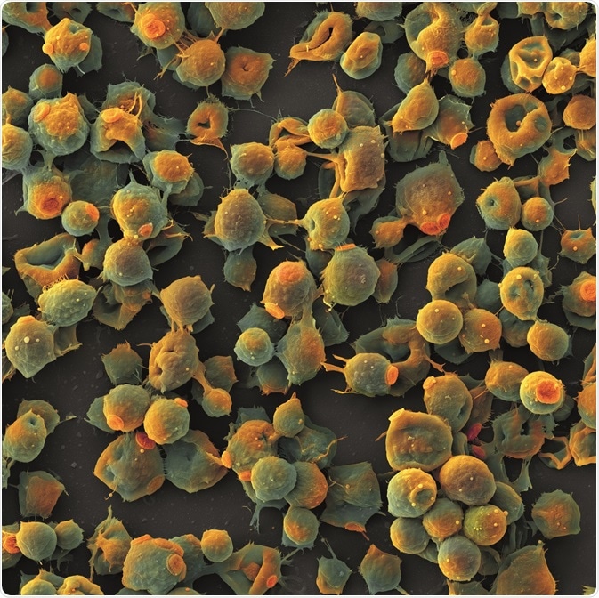
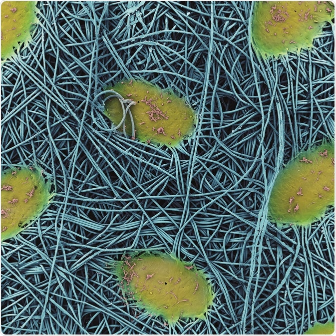
Left: Brettanomyces – Brettanomyces yeast cells that are responsible for the ‘Brett’ character of wine. Right: Filtr – Pores of a membrane filter. This type of microfilter with pore sizes from 0.45 to 1.2µm is used to purify wine immediately before bottling.
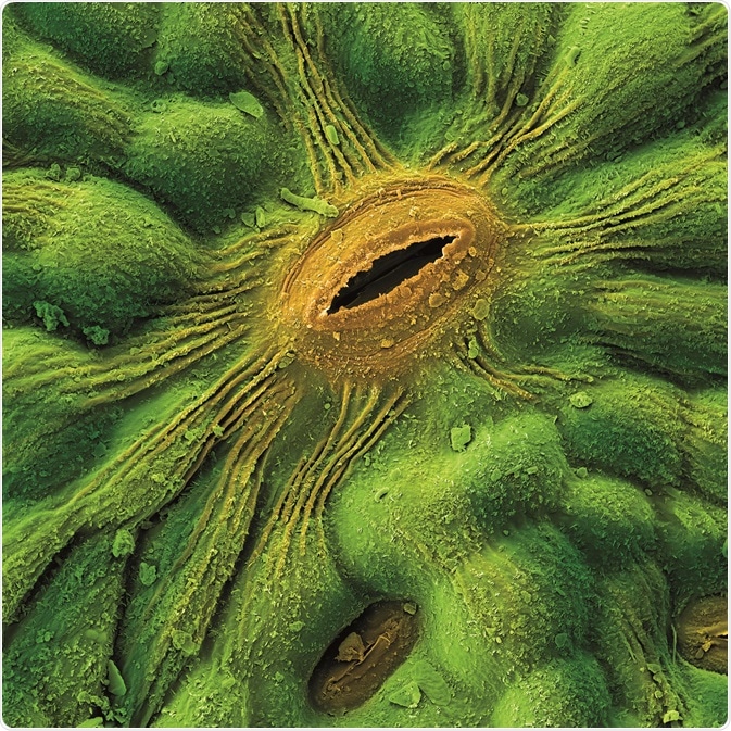
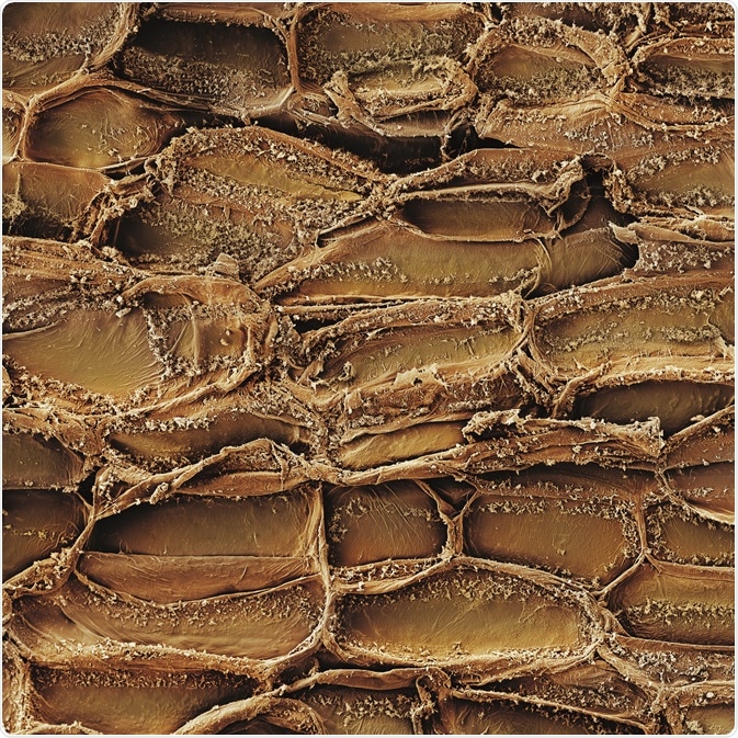
Left: Grape leaf – Details of a stoma, a respiration pore located on the underside of a grape leaf. Stomata are used for nutrient exchange (e.g. carbon dioxide and oxygen) between the plant and surroundings. Right: Kura – Similar to human skin, bark is the dead tissue on the outer layer of a tree. It serves to protect the tree from excessive water loss, regulates gas exchange and excretes some metabolites. Shown here are dead epidermis tissue cells.
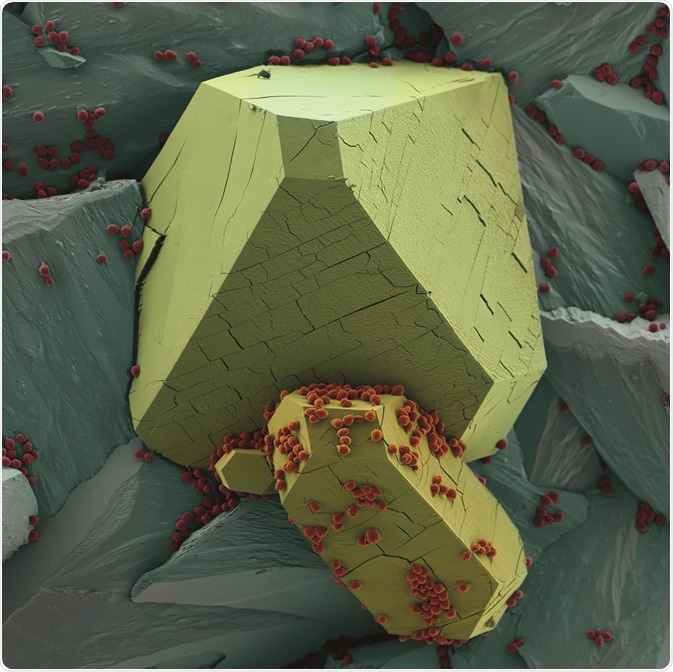
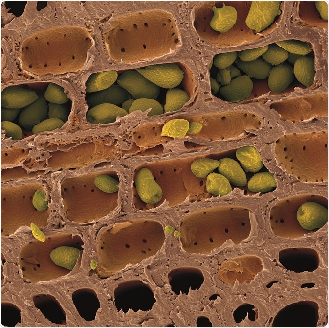
Left: Vinny Kamen – Potassium bitartrate or potassium hydrogen tartrate is potassium salt of tartaric acid present in wine. Crystallization of this compound leads to turbidity in the form of fine sediment. Shown here is potassium bitartrate with yeast cells on the surface. Right: Wood – Vascular tissue transport organic and inorganic substances around the plant. Shown here are starch grains deposited in vascular tissues of a grapevine.
Last updated: Mar 27, 2019 at 5:36 AM


No hay comentarios:
Publicar un comentario