
NIGMS Is on Instagram!
NIGMS is now on Instagram (@NIGMS_NIH), beaming all the gorgeous science images you can’t get enough of straight to your mobile devices
NIGMS Is on Instagram!
Science is beautiful.
For several years, we’ve used this blog to highlight pictures we think are cool, scientifically relevant and visually striking. The images were created by NIGMS-funded researchers in the process of doing their research. Many come from our Life: Magnified collection, which features dozens of stunning photos of life, close-up. We’ll continue to bring you interesting images and information here on Biomedical Beat, but if you can’t get enough of them, we have a new way to share our visual content with you: Instagram.
We’re pleased to announce the launch of our NIGMS Instagram account. We’ll highlight gorgeous images, and bring you the science behind them—straight from our mobile device to yours. Instagram lets us label our images with subject-specific hashtags. You can find our pictures by going on Instagram and searching for #NIGMS. If you’re already an Instagram user, you can follow us @NIGMS_NIH. You can even see our page using a web browser at https://www.instagram.com/nigms_nih/  . Let us know what you think!
Also, if you have any stunning images or videos that relate to scientific areas supported by NIGMS, please send them to us. They might end up on our Instagram feed!

See those finger-like projections? They are called villi. This image shows the small intestine, where most of the nutrients from the food we eat are absorbed into the bloodstream. The villi increase the organ’s surface area, making nutrient absorption more efficient. Credit: National Center for Microscopy and Imaging Research.
#science #biology #research #cells #cellbiology #microscopy #nigms #scienceisbeautiful #sciart #nigms #nih #NCMIR #UCSD #smallbowel #nutrition #nutrients #nutrient #anatomyphysiology
#science
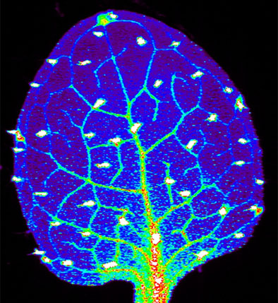
We need zinc. It’s an essential nutrient for growth and development, fending off invading microbes, healing injuries, and all sorts of cellular processes. We get the mineral through our diet, but people in certain parts of the world don’t get enough. Researchers study how plants acquire and process zinc, hoping to find ways to increase the nutrient in food crops. Using synchrotron X-ray fluorescence technology, scientists created this heat map of zinc in a leaf from a plant called Arabidopsis thaliana (zinc levels from lowest to highest: blue, green, red, white). Credit: Suzana Car and Mary Lou Guerinot, Dartmouth College.
#science #biology #research #botany #plant #arabidopsis #zinc #microscopy #synchrotron #x-ray #modelorganism #leaf #heatmap #scienceisbeautiful #nigms #nih
#science
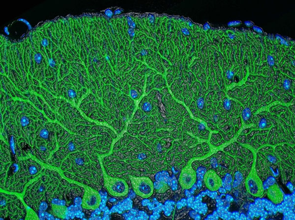
Beautiful brain! This image shows the cerebellum, which is the brain’s locomotion control center. Every time you shoot a basketball, tie your shoe or chop an onion, your cerebellum fires into action. Found at the base of your brain, the cerebellum is a single layer of tissue with deep folds like an accordion. People with damage to this region of the brain often have difficulty with balance, coordination and fine motor skills. Credit: Tom Deerinck and Mark Ellisman, NCMIR
#science #biology #research #cerebellum #neuroscience #neurobiology #brain #brains #brainimages #neuroanatomy #microscopy #scienceisbeautiful #sciart #nigms #nih #NCMIR #UCSD
#science
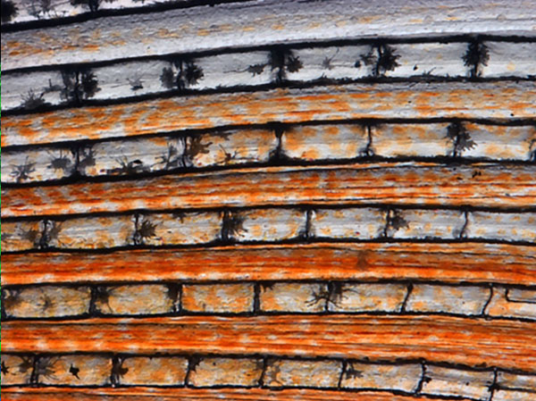
Scientists can learn a lot by studying pigment cells, which give animals their colorful skins, eyes, hair and scales. We can even gain insight into skin cancers, like melanoma, that originate from pigment cells. Pigment cells can form all sorts of patterns, like these stripes on the fin of a pearl danio, a type of tropical minnow. Credit: David Parichy, University of Washington.
#biology #marinebiology #marinescience #cell #pigmentcell #xanthophore #science #research #cellbiology #microscopy #scienceisbeautiful #scienceiscool #zebrafish #universityofwashington
#biology


.png)


No hay comentarios:
Publicar un comentario