Volume 26, Number 9—September 2020
Research Letter
Effect of Environmental Conditions on SARS-CoV-2 Stability in Human Nasal Mucus and Sputum
On This Page
Figures
Tables
Downloads
Article Metrics
M. Jeremiah Matson, Claude Kwe Yinda, Stephanie N. Seifert, Trenton Bushmaker, Robert J. Fischer, Neeltje van Doremalen, James O. Lloyd-Smith, and Vincent J. Munster
Abstract
We found that environmental conditions affect the stability of severe acute respiratory syndrome coronavirus 2 in nasal mucus and sputum. The virus is more stable at low-temperature and low-humidity conditions, whereas warmer temperature and higher humidity shortened half-life. Although infectious virus was undetectable after 48 hours, viral RNA remained detectable for 7 days.
Severe acute respiratory syndrome coronavirus 2 (SARS-CoV-2) is shed predominantly in upper and lower airway secretions (1), and transmission likely occurs predominantly through respiratory droplets, and potentially through direct contact and fomites. We describe SARS-CoV-2 stability in human nasal mucus and sputum under different environmental conditions.
We acquired pooled human nasal mucus and sputum commercially (Lee BioSolutions Inc., ) and mixed it with SARS-CoV-2 (SARS-CoV-2/human/USA/USA-WA1/2020) (2). We aliquoted 50 μL of each fluid containing 1 × 10⁵ 50% tissue culture infective dose/mL SARS-CoV-2 into sealed tubes (liquid setting) or onto polypropylene disks (surface setting), as previously described (3). We assessed stability under 3 environmental conditions: 4°C/40% relative humidity (RH), 21°C/40% RH, and 27°C/85% RH (RH applies only to exposed surface samples). We collected samples at specified timepoints and analyzed them for infectious virus by using endpoint titration. We extracted aliquots of collected surface samples by using the QIAGEN QIAamp Viral RNA Mini Kit (QIAGEN, ) and analyzed them for the presence of viral RNA by using a quantitative reverse transcription PCR assay targeting the E gene (4). We fit linear regression models to log10-transformed titer data, calculated SARS-CoV-2 half-life (t1/2) for each condition, and tested differences by using analysis of covariance. We report all experimental measurements as means of 3 replicates with SE. We considered differences with p values <0.05 statistically significant.
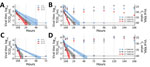
Figure. Stability of severe acute respiratory syndrome coronavirus 2 over time in human nasal mucus and sputum under different environmental conditions: liquid nasal mucus (A), surface nasal mucus (B), liquid sputum (C),...
We observed no significant differences in SARS-CoV-2 t1/2 between environmental conditions in liquid nasal mucus. In surface nasal mucus, SARS-CoV-2 t1/2 was significantly shorter at 27°C/85% RH compared with 21°C/40% RH (p = 0.0023) and 4°C/40% RH (p = 0.0007). At 27°C/85% RH, SARS-CoV-2 t1/2 also was significantly shorter in surface compared with liquid nasal mucus (p = 0.0101). Other comparisons of nasal mucus did not demonstrate significant differences in SARS-CoV-2 t1/2 (Table; Figure, panel A, B).
SARS-CoV-2 t1/2 was significantly longer in liquid sputum at 4°C than at 21°C (p = 0.0006) and 27°C (p<0.0001). In surface sputum, SARS-CoV-2 t1/2 also was significantly longer at 4°C/40% RH than at 21°C/40% RH (p = 0.0042) and 27°C/85% RH (p = 0.0002). In addition, SARS-CoV-2 t1/2 was significantly longer at 21°C/40% RH than 27°C/85% RH (p = 0.0027) in surface sputum. We observed no significant differences in SARS-CoV-2 t1/2 between liquid and surface sputum (Table; Figure, panel C, D). SARS-CoV-2 RNA remained detectable for >7 days in all surface samples, with a gradual increase in cycle threshold value (decrease in detected RNA) occurring only in nasal mucus at 27°C/85% RH (Figure, panel B, D).
We previously reported on the surface stability of SARS-CoV-2 in culture media at 21°C/40% RH (3). However, SARS-CoV-2 stability is affected by its surrounding matrix and environmental conditions. The t1/2 we report here for SARS-CoV-2 in surface nasal mucus and sputum at 21°C/40% (Table) is considerably shorter than what we found in culture media under similar conditions (t1/2 6.8 [95% CI 5.6–8.2] hours) (3). In addition, we set out to determine SARS-CoV-2 stability in nasal mucus and sputum under environmental conditions that approximate temperate winter (4°C/40% RH), climate-controlled (21°C/40% RH), and temperate summer or tropical (27°C/85% RH) settings. SARS-CoV-2 was generally more stable at cooler temperatures and lower RH, and less stable at warmer temperatures and higher RH. Nevertheless, with our experimental protocol and initial titer, we predicted that SARS-CoV-2 would remain infectious in nasal mucus and sputum on surfaces for >10–12 hours even in warm, humid conditions. However, the amount of infectious SARS-CoV-2 shed and virus stability in relationship to infectious dose for humans are currently unknown, hampering conclusions regarding infectious duration and transmission. The general similarity in SARS-CoV-2 stability between liquid and surface samples suggests that in general temperature factored more heavily than humidity.
Community transmission of SARS-CoV-2 is widespread (5) and might be explained by contact with asymptomatic or presymptomatic (6) infected persons. Because of the surface stability of SARS-CoV-2, fomite transmission might also play a role. In addition, reduced surface stability of SARS-CoV-2 in human nasal mucus and sputum in warmer and more humid conditions might result in decreased virus transmission, and climatic influence on SARS-CoV-2 transmission rates might eventually drive seasonal outbreak dynamics in a postpandemic period (7), similar to other respiratory viruses (e.g., influenza A virus or human coronavirus OC43).
SARS-CoV-2 RNA has been detected on surfaces throughout clinical settings (8,9) and aboard a cruise ship for extended periods (10), but any correlation to infectious virus was previously unknown. In our study, infectious virus persisted in both nasal mucus and sputum on surfaces for ≈24 hours under climate-controlled conditions. However, viral RNA was consistently detectable for >7 days under various conditions in both nasal mucus and sputum on surfaces. These findings suggest that inferences regarding the presence of infectious virus from quantitative reverse transcription PCR data alone should be made with caution.
Mr. Matson is an MD/PhD student at the Marshall University Joan C. Edwards School of Medicine and is performing his dissertation research at the National Institute of Allergy and Infectious Diseases Virus Ecology Section. He is interested in emerging and reemerging viruses.
Acknowledgments
We thank Emmie de Wit, Brandi Williamson, and Jon Schulz for their technical assistance.
This research was supported by the Intramural Research Program of the National Institutes of Health, the National Institute of Allergy and Infectious Diseases, and the Defense Advanced Research Projects Agency’s Preventing Emerging Pathogenic Threats Program (grant no D18AC00031).
References
- Zhu N, Zhang D, Wang W, Li X, Yang B, Song J, et al.; China Novel Coronavirus Investigating and Research Team. A novel coronavirus from patients with pneumonia in China, 2019. N Engl J Med. 2020;382:727–33.
- Harcourt J, Tamin A, Lu X, Kamili S, Sakthivel SK, Murray J, et al. Severe acute respiratory syndrome coronavirus 2 from patient with coronavirus disease, United States. Emerg Infect Dis. 2020;26:1266–73.
- van Doremalen N, Bushmaker T, Morris DH, Holbrook MG, Gamble A, Williamson BN, et al. Aerosol and surface stability of SARS-CoV-2 as compared with SARS-CoV-1. N Engl J Med. 2020;382:1564–7.
- Corman VM, Landt O, Kaiser M, Molenkamp R, Meijer A, Chu DK, et al. Detection of 2019 novel coronavirus (2019-nCoV) by real-time RT-PCR. Euro Surveill. 2020;25:25.
- Liu J, Liao X, Qian S, Yuan J, Wang F, Liu Y, et al. Community transmission of severe acute respiratory syndrome coronavirus 2, Shenzhen, China, 2020. Emerg Infect Dis. 2020;26:1320–3.
- He X, Lau EHY, Wu P, Deng X, Wang J, Hao X, et al. Temporal dynamics in viral shedding and transmissibility of COVID-19. Nat Med. 2020;26:672–5.
- Kissler SM, Tedijanto C, Goldstein E, Grad YH, Lipsitch M. Projecting the transmission dynamics of SARS-CoV-2 through the postpandemic period. Science. 2020;368:860–8.
- Ong SWX, Tan YK, Chia PY, Lee TH, Ng OT, Wong MSY, et al. Air, surface environmental, and personal protective equipment contamination by severe acute respiratory syndrome coronavirus 2 (SARS-CoV-2) from a symptomatic patient. JAMA. 2020;323:1610.
- Guo ZD, Wang ZY, Zhang SF, Li X, Li L, Li C, et al. Aerosol and surface distribution of severe acute respiratory syndrome coronavirus 2 in hospital wards, Wuhan, China, 2020. Emerg Infect Dis. 2020;26:26.
- Moriarty LF, Plucinski MM, Marston BJ, Kurbatova EV, Knust B, Murray EL, et al.; CDC Cruise Ship Response Team; California Department of Public Health COVID-19 Team; Solano County COVID-19 Team. California Department of Public Health COVID-19 Team; Solano County COVID-19 Team. Public health responses to COVID-19 outbreaks on cruise ships—worldwide, February–March 2020. MMWR Morb Mortal Wkly Rep. 2020;69:347–52.
Figure
Table
Suggested citation for this article: Matson MJ, Kwe Yinda C, Seifert SN, Bushmaker T, Fischer RJ, van Doremalen N, et al. Effect of environmental conditions on SARS-CoV-2 stability in human nasal mucus and sputum Emerg Infect Dis. 2020 Sep [date cited]. https://doi.org/10.3201/eid2609.202267
Original Publication Date: June 08, 2020


.png)


.png)


No hay comentarios:
Publicar un comentario