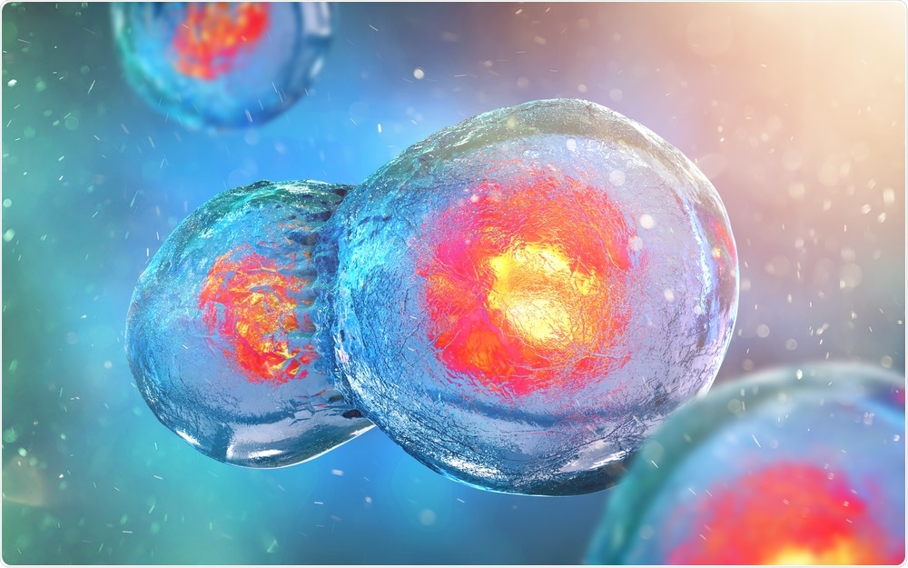
Researchers develop the first interactive model of cell division
Scientists from the European Molecular Biology Laboratory (EMBL) have created the first interactive map of the proteins involved in cell division.
 Image Credit: Andrii Vodolazhskyi / Shutterstock
The tool enables researchers to pinpoint which proteins work together to drive this essential process. The importance of this is paramount, as the failure of mitosis leads to defects such as fertility problems and cancer.
In 2010, the same research team established which parts of the human genome are involved in cell division, but it is not the genome that drives the process; it is the proteins the genome encodes.
Hundreds of proteins work together to coordinate cell division, often working in groups, but until now, researchers have mainly focused on single proteins.
Project leader Jan Ellenberg says that now it is possible to take a “systems approach” and study the bigger picture by looking at the dynamic networks the proteins form in living human cells.
The Mitotic Cell Atlas integrates this information into an interactive 4D computer model that researchers can use to select any combination of mitotic proteins and see how they cooperate during cell division.
Besides mitosis, the technologies developed here can be used to study proteins that drive other cellular functions, for example cell death, cell migration or metastasis of cancer cells.
Overall, about 600 proteins drive mitosis in human cells. Having a dataset for all 600 of them would enable researchers to understand how information is transmitted and how decisions are made within a cell as it divides. This will require several more years of research.
Co-author Stephanie Alexander says that at EMBL, the researchers are continuously adding data to the atlas by imaging more and more proteins using the same standardized method:
Source:
This news article has been re-written from an EMBL press release, originally published on EurekAlert.


No hay comentarios:
Publicar un comentario