Advanced live cell imaging reveals brain’s response to blood vessel injury
Live cell imaging and laser beam ablation reveals the damage response of pericytes surrounding blood vessels in the brain.
When blood vessels in the brain are injured, cells present on the vessels called pericytes, extend into the space left when nearby pericytes die to compensate for their loss, according to a study conducted at the Medical University of South Carolina (MUSC).
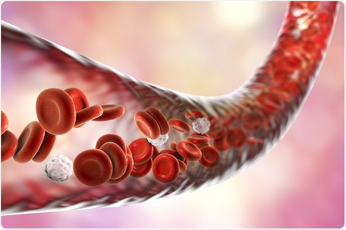
 Credit: Kateryna Kon/Shutterstock.com
Principal investigator Andy Shih and colleagues from the MUSC Department of Neuroscience are keen to find out whether this compensatory growth, which is a form of brain plasticity, could be manipulated to combat conditions such as stroke and Alzheimer’s disease, which are characterized by a high degree of pericyte loss.
Pericytes aid the maturation of blood vessels in the brain as it grows, but little is understood about their function in the adult brain and why these cells seem to be so vulnerable to injury in stroke, Alzheimer’s disease and other brain disorders.
For the study, live adult mice were genetically modified so that their brain pericytes could be strongly illuminated under a two-photon microscope. This advanced imaging technique revealed the pericytes to be oval cell bodies, often positioned near junctions where two capillaries intersected, with tentacle-like arms (processes) extending outwards along the capillaries. The tips of those arms seemed to almost touch the arm tips of neighboring pericytes, thereby creating a “blanket” to cover the capillaries.
To investigate what happens when pericytes are lost, Shih and team ablated one pericyte at a time using a high-precision laser beam. When a pericyte was lost, the capillary it had been covering seemed to dilate.
The researchers continued to ablate more pericytes and over a period of days to weeks, the processes of nearby pericytes grew to cover the exposed capillaries, restoring their normal tone and dilation. Maintenance of this capillary tone is essential to keeping blood flow in the brain healthy.
The images of pericytes extending their arms to cover exposed capillaries are the first of their kind says Shih: "Most methods to study pericytes give you a snapshot in time. To be able to do this dynamically in a live animal's brain is an important step forward.”
However, Shih suspects there is a limit to how much pericytes can grow to cover exposed capillaries and it is still not clear what happens when larger numbers of pericytes die, as is the case in stroke and Alzheimer's disease. The team’s next study, which will be funded by The Alzheimer’s Association, will look at blood vessel health when large numbers of pericytes are ablated.
"Are there ways to augment this plasticity, to protect it and stabilize it if we need to? There are mechanisms driving this that we need to understand," says Shih.


.png)


No hay comentarios:
Publicar un comentario