Mimicking Human Brain Development Using Organoids
 Thought LeadersDr. Paul TesarTESAR LaboratoryCase Western Reserve University
Dr. Paul Tesar, D.Phil., from the TESAR Laboratory at CWRU School of Medicine, Ohio, discusses the importance of organoids in biological research and the development of organoids which are capable of simulating the early stages of human myelin.
What are organoids? Why are they useful for studying human development?
Organoids are three-dimensional pieces of tissue generated in a laboratory that exhibit simple features, functions and organized structure of an organ. The growth of organoids in the laboratory typically begins with human embryonic stem cells or induced pluripotent stem (iPS) cells.
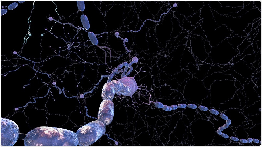
 Image Credit: 3Dme Creative Studio / Shutterstock
The development of human iPS cells in particular means we now have the potential to create organoids from any human subject, including those afflicted with certain diseases.
The key advantage of organoid technology is that it provides the ability to generate scalable quantities of developing human tissue in the lab, which was previously impossible, especially in the context of the human brain.
Are there any limitations when it comes to using organoids to study the human brain?
There are several pioneers that have led both the discovery as well as recent advances in human brain organoid technology.
Even though this technology is very powerful and offers a tremendous amount of potential, most scientists would agree that we are still in the beta phase and that a number of sequential advancements will be necessary for us to create organoids that provide a more realistic representation of human brain tissue.
Currently, we are only able to model certain aspects of early human brain development with this organoid technology, as we are limited by the simplistic organization and lack of maturation within these organoids.
Why is it important that new methods of producing organoids are developed?
The organoid field is advancing at a very rapid pace. Early pioneering technologies largely consisted of self-organizing neural progenitor cells and neurons.
Whereas now, we have organoids that can mature to include contain additional cell types such as astrocytes and vasculature, which enables a more accurate representation of the complex three-dimensional native brain tissue.
However, more still need to be done to achieve organoids that contain the full repertoire of cell types normally seen in fully functioning brain tissue.
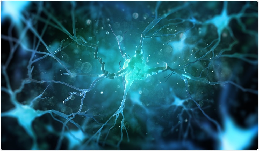
 Image Credit: Andrii Vodolazhskyi / Shutterstock
Please describe your recent research.
My laboratory is focused on understanding the development of human oligodendrocytes and their dysfunction in neurological disorders like multiple sclerosis and pediatric leukodystrophies such as Pelizaeus-Merzbacher disease.
Along with astrocytes and neurons, oligodendrocytes are one of the three major cell types in the central nervous system. These cells are responsible for creating myelin, a discontinuous lipid-based substance that coats neuronal axons.
Without myelin, neurons fail to appropriately conduct electrical signals, leading to neural dysfunction and disability.
In the organoid field, we have whole brain organoids and spheroids, which represent a more restricted sub-region of the tissue such as the cortex, in our case.
Organoid and spheroid technologies provide a powerful platform from which to examine oligodendrocytes and myelin biology, but thus far, organoids have lacked oligodendrocytes and myelin.
This got us thinking, how can we produce organoids that contain these essential components? We modified previous methods developed by pioneers in the field of organoid technology in in a way that allowed us to stimulate the generation and maturation of oligodendrocytes within induced pluripotent stem cell-derived organoids.
We call these oligocortical spheroids. These spheroids are capable of simulating the early stages of human myelin, which is one of the first examples of access to the stages of human myelination in the laboratory.
How will this research impact patients with Pelizaeus-Merzbacher disease?
This research should have a huge impact on our understanding of Pelizaeus-Merzbacher disease. Pelizaeus-Merzbacher disease is a rare X-linked myelin disorder that typically affects young children. It is caused by mutations in the PLP1 gene and has a range of clinical effects, depending on the mutations involved.
So far, we have not been able to fully understand how each of these mutations causes myelin dysfunction in patients. However, using these oligocortical spheroids, we are now able to produce brain-tissue or tissue-like material from iPS cells generated from patient samples.
This will allow us to study the temporal, cellular, and molecular pathological underpinnings of Pelizaeus-Merzbacher disease.
More importantly, it will allow us to potentially screen or develop therapeutics that can modulate the disease pathology. This could have the potential to restore myelin function to Pelizaeus-Merzbacher patients in the future.
Using this method, can we create models for any neurological condition? Are there any drawbacks to the technique?
While oligocortical steroids offer a tremendous amount of potential for understanding human brain development and function, it is still in the early stages of development.
Currently, the technology can provide some key insights into the human brain, but in order to be able to model the more complex pathology of adult-onset disorders and normal tissue homeostasis in the adult brain, new methods will be needed.
We need to be able to generate organoids that can mature enough to reflect the later stages of human development and be maintained in the laboratory for an extended period. This will allow us to understand normal human physiology and compare this to the pathology of human diseases. Currently, we are following both pathways of advancing the technology and using the technology as it stands to give us novel insight.
What does the future hold for your research?
In general, my laboratory functions at the interface of neuroscience and stem cell technology. We strive to develop cellular tools that can further our understanding of neural development and disease.
When it comes to oligocortical steroids, we are working to further refine and optimize this technology to reflect more mature stages of human myelination.
Another goal of ours is to be able to model other regions of the central nervous system outside of the cortex, which is where our previous studies have been focused.
Finally, as previously mentioned, we are working to use this oligocortical steroid platform to help identify potential therapeutics that can enhance the generation of human oligodendrocytes and human myelin, both in the context of disease like multiple sclerosis, but also in the context of genetic disorders, including Pelizaeus-Merzbacher disease.
Where can readers find more information?
About Dr. Paul Tesar
 Dr. Paul Tesar is currently an Associate Professor and the Dr. Donald and Ruth Weber Goodman Professor of Innovative Therapeutics at CWRU School of Medicine in the Department of Genetics and Genome Sciences.
After receiving his undergraduate degree in biology from Case Western Reserve University, Paul went on to earn his D.Phil. (Ph.D.) from the University of Oxford as a recipient of a prestigious scholarship from the National Institutes of Health.
Paul’s scientific achievements continue to be recognized with a number of prestigious awards including being named a Robertson Investigator of the New York Stem Cell Foundation in 2011.
One of only four international awardees, the honor recognizes and supports scientists leading their generation in stem cell research.
Paul also co-founded a Cleveland-based biotechnology company to advance new therapies from the laboratory into clinical testing to better the lives of patients and their families.


No hay comentarios:
Publicar un comentario