|
|
| | September 26, 2018 | |
| | |
| | The latest life science microscopy news from AZoNetwork | |
|
|
 |
| |  Combination of Technologies Drives Advances in Life Science Imaging Combination of Technologies Drives Advances in Life Science Imaging
Over the years, Bruker have developed a life science microscopy business that specializes in advanced technologies for neuroscience, live-cell imaging, and molecular imaging.
Now, with the acquisition of JPK Instruments, Bruker's portfolio will be further augmented by JPK’s advanced technologies and applications. JPK adds in-depth expertise in live-cell imaging, cellular mechanics, adhesion, and molecular force measurements, optical trapping, and biological stimulus-response characterization to Bruker.
| |
|
|
|
|
|
|
| |
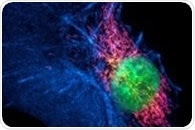
 In a revolutionary study, an LMU research team headed by Ralf Jungmann has shown that the application of chemically-modified DNA aptamers as protein markers enables one to improve the power of super-resolution fluorescence microscopy as an imaging instrument. In a revolutionary study, an LMU research team headed by Ralf Jungmann has shown that the application of chemically-modified DNA aptamers as protein markers enables one to improve the power of super-resolution fluorescence microscopy as an imaging instrument. | |
|
| |
 Despite being produced decades ago, the use of complementary metal oxide semiconductor (CMOS) image sensors in optical microscopy was often overlooked. In this article, we look at how CMOS image sensors have started to be used in some optical microscopes. Despite being produced decades ago, the use of complementary metal oxide semiconductor (CMOS) image sensors in optical microscopy was often overlooked. In this article, we look at how CMOS image sensors have started to be used in some optical microscopes. | |
|
| |
 Photoactivated Localization Microscopy (PALM) is a form of super resolution fluorescence microscopy (SR) which enables highly resolved imaging to be produced through the selective targeting of fluorescent markers within a specimen. Photoactivated Localization Microscopy (PALM) is a form of super resolution fluorescence microscopy (SR) which enables highly resolved imaging to be produced through the selective targeting of fluorescent markers within a specimen. | |
|
| |
 Structured illumination microscopy (SIM) is a form of super-resolution light microscopy that enhances the resolution of the microscope. It is useful in research as it allows for high-resolution imaging of cells. Structured illumination microscopy (SIM) is a form of super-resolution light microscopy that enhances the resolution of the microscope. It is useful in research as it allows for high-resolution imaging of cells. | |
|
| |
 Scattering-Assisted Localization Microscopy (SALM) is a super-resolution microscopy tool where high resolution is obtained based on the size of the grain in the speckle pattern on the sample. Scattering-Assisted Localization Microscopy (SALM) is a super-resolution microscopy tool where high resolution is obtained based on the size of the grain in the speckle pattern on the sample. | |
|
|
|
|
|


.png)


No hay comentarios:
Publicar un comentario