Miniaturized Device Puts Nanotechnology to Work in Diagnosing Cancer
An experimental diagnostic device could be a valuable addition to a pathologist’s tool kit for diagnosing cancer, new research suggests. The miniaturized nuclear magnetic resonance, or micro-NMR, device can diagnose cancer within an hour, using patient samples of just a few thousand cells that are collected using a fine needle and a syringe.
Initial clinical studies by researchers at Massachusetts General Hospital (MGH) indicate that the micro-NMR system, which can quantify multiple protein markers in cells from a single patient sample, may be more accurate than standard diagnostic techniques.
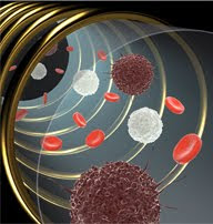
A miniaturized nuclear magnetic resonance (NMR) device can detect cancer cells (dark brown) in a small sample of cells from a patient. The coils generate magnetic fields that excite magnetic nanoparticles attached to antibody-protein complexes, resulting in NMR signals that can be used to identify a cancer protein signature. (Image courtesy of H. Lee and R. Weissleder, Massachusetts General Hospital)
Conventional pathology, which is the current gold standard for diagnosing many cancers, is far from perfect. It takes several days to yield results, may require surgery to provide enough tissue, is technically challenging, and is subject to human interpretation. A biopsy for conventional pathology may be done either with open surgery, which yields samples containing billions of cells, or with a wide or fine needle. A pathologist processes and stains the tissue sample and examines it under a microscope. If enough tissue is available, the pathologist may use immunohistochemistry to visualize specific cancer markers in cross-sections of tissue.
The micro-NMR device uses principles similar to those of magnetic resonance imaging (MRI) to detect magnetic nanoparticles attached to antibodies, which flag protein biomarkers known to be associated with some cancers in patient samples. A physician can operate the portable device from the patient’s bedside with a smart-phone application that displays results on the phone’s screen.
Simplicity, Speed, and Sensitivity
The reactions for coupling the magnetic nanoparticle-antibody complexes to specific target proteins are performed at room temperature, and sample processing is simple and quick, the researchers said. In addition to eliminating waiting time, the rapid processing is an advantage because experiments showed that protein markers in patient samples degrade quickly.
Micro-NMR probably won’t replace conventional pathology and histology completely, but “it would be a good mechanism to triage patients before they undergo more invasive procedures,” said Dr. Cesar Castro, an oncologist and investigator at MGH. Dr. Castro co-led a recent clinical study that was the first to test the micro-NMR and nanoparticle technologies in patient samples.
The entire micro-NMR system costs about $200 and is roughly the size of a cube-shaped box of facial tissue. Developed by Drs. Hakho Lee and Ralph Weissleder at the Center for Systems Biology at MGH, the system is highly sensitive thanks to miniaturization of the device and the chemistry used to bind magnetic nanoparticles to their targets. The device can measure levels of individual tumor markers in a 1-microliter sample containing about 200 cells and—unlike technologies that rely on optical properties—works in nonpurified samples.
Advantages in the Clinic
If validated in larger clinical trials, the micro-NMR system could eventually be used not only to diagnose cancer but also to identify potential patient candidates for targeted therapies and to monitor the response to therapy, Dr. Castro said. When used for diagnosis, this approach could potentially reduce the time that patients are in limbo while waiting for results and provide more information to clinicians, while steering patients away from more invasive procedures and avoiding repeat biopsies, he noted.
“A key advantage of this technique is the ability to look at multiple markers at the same time,” said Dr. Piotr Grodzinski, director of NCI’s Office of Cancer Nanotechnology Research, which helped fund the work. This so-called multiplexing capability is important, he explained, “because cancer is heterogeneous and is not characterized by a single biomarker.”
The diagnostic micro-NMR system includes a mini magnet (left); chip-sized, microliter-volume sensors (center); and a user-friendly smart phone interface (right). (Image courtesy of C. Min, D. Issadore, R. Weissleder and H. Lee)
The MGH researchers used micro-NMR to measure levels of nine established cancer-related markers in cells from fine-needle biopsies of deeper lesions, done with guidance from a CT scan or ultrasound. Using a four-protein signature, they were able to diagnose a range of epithelial cancers, including lung, breast, pancreatic, and gastrointestinal, with 96 percent accuracy.
In cell samples from 50 patients who had been referred for clinical biopsies, the four-marker panel correctly diagnosed 48 cases: 44 of 44 malignancies and 4 of 6 benign lesions. In contrast, the accuracy of conventional cytology and histology performed on specimens from the same patients was 74 percent and 84 percent, respectively. The diagnoses were confirmed independently using a combination of clinical, imaging, and pathology data.
The next step for Dr. Castro and his colleagues is to refine the system to identify specific cancers. “Our first endeavor will be customizing the assay with ovarian cancer-specific markers, and we’re enrolling patients treated at the MGH Gillette Center for Gynecologic Oncology for that study,” Dr. Castro said. The researchers hope eventually to apply the technology to blood and other fluids, such as ascites, in which tumor cells are scarce, and they are exploring other cancer applications as well.
Potential for Personalized Medicine
Many cancer researchers and companies recognize the need for diagnostic tools that could be used in tandem with targeted therapies, and in a range of clinical studies needed to develop such therapies.
“To fully achieve the potential of personalized cancer therapy will require noninvasive methods for analyzing genes and proteins in a patient’s tumor or serum, which can be used in real time to determine the best treatment or treatment combination,” said Dr. Roy Herbst, who recently assumed the role of chief of medical oncology at Yale Medical Center. “I think this [micro-NMR system] is clearly an example of a step in the right direction.”
“With this machine, we have the strong potential to start asking more refined and elegant questions in clinical research,” Dr. Castro said. Because the micro-NMR technology is sensitive and minimally invasive, he explained, it could be used in clinical trials that require repeated tumor sampling at various time points. For example, researchers could use micro-NMR to identify and validate new tumor markers for clinical use or to look at how markers change during the course of therapy.
“We consider this to be a platform technology because the markers are very interchangeable,” Dr. Castro explained. “So, as the science evolves, as we get more information from the clinic and from laboratories elsewhere, we can react to that.”
—Elia Ben-Ari
Biography - Elia Ben-Ari
After a brief stint in biomedical research, Elia Ben-Ari left the lab bench in 1991 to pursue a career as a science writer and editor, working in journalism, publishing, and public affairs. She enjoys learning and writing about diverse topics in biology and medicine ranging from molecules to ecosystems, and is also interested in the intersection of science and the humanities. Elia joined the Bulletin staff in 2010.
full-text:
NCI Cancer Bulletin for March 22, 2011 - National Cancer Institute
Suscribirse a:
Enviar comentarios (Atom)


.jpg)


No hay comentarios:
Publicar un comentario