|
|
| | March 14, 2019 | |
| | |
| | The latest life science microscopy news from AZoNetwork | |
|
|
 |
| |  Small Molecules, Big Challenges: Adopting New Methods for Stability Testing Small Molecules, Big Challenges: Adopting New Methods for Stability Testing
A technique that enables determination of active ingredient content of tablets during continuous manufacturing processes is Raman spectroscopy. In addition, this has been combined with optical imaging to form the methodology known as Raman microscopy. This is a powerful tool for providing detailed chemical information in both the spatial and spectral dimensions. As such, it has become the standard way for the pharmaceutical industry to achieve polymorphic characterization of active pharmaceutical ingredients.
| |
|
|
|
|
|
|
| |
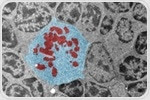
 Two-photon microscopy is a method that enables the imaging of live cell and tissue samples with high resolution. It is a form of fluorescence microscopy and provides visualization of fluorescent signals via an excitation wavelength being absorbed by a fluorophore and an emission wavelength released. Two-photon microscopy is a method that enables the imaging of live cell and tissue samples with high resolution. It is a form of fluorescence microscopy and provides visualization of fluorescent signals via an excitation wavelength being absorbed by a fluorophore and an emission wavelength released. | |
|
| |
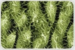 There are several different types of scanning probe microscopes, the most prominent of which are atomic force microscopy (AFM) and scanning tunneling microscopy (STM). There are also many other types, which are listed at the end of this article. There are several different types of scanning probe microscopes, the most prominent of which are atomic force microscopy (AFM) and scanning tunneling microscopy (STM). There are also many other types, which are listed at the end of this article. | |
|
| |
 This British Science Week, we are celebrating the scientific pioneers of the past, present and future. Perhaps one of the greatest British pioneers in microscopy was Robert Hooke, who was the first person to observe insect and plant cells using various magnifying lense. This British Science Week, we are celebrating the scientific pioneers of the past, present and future. Perhaps one of the greatest British pioneers in microscopy was Robert Hooke, who was the first person to observe insect and plant cells using various magnifying lense. | |
|
|
|
|
|


.png)


No hay comentarios:
Publicar un comentario